


Case Presentation:
Carotid Ophthalmic Aneurysm - Case 1
History and Physical
- 48-year-old lady presented to the emergency room with severe headache. Subsequent work-up demonstrated subarachnoid hemorrhage and she underwent cerebral angiography.
- Her neurological examination was non focal.
Imaging
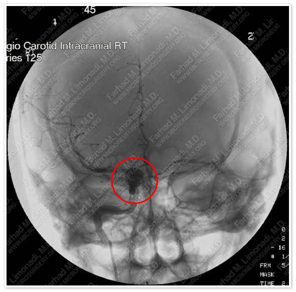
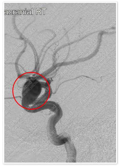
Cerebral angiography of the patient’s brain shows a carotid ophthalmic artery communicating artery aneurysm on the right.
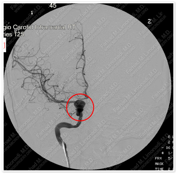
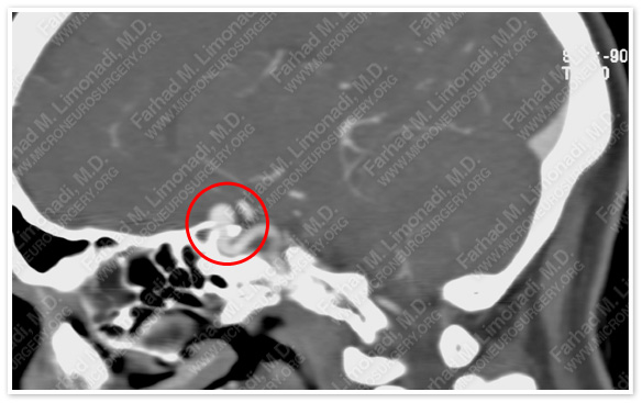
Post-op Imaging
Before Operation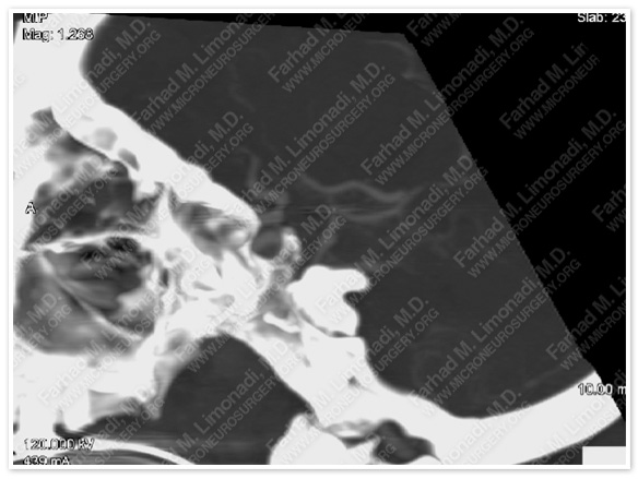
After Operation
Post-op CT angiography shows exclusion of the dome of this complex aneurysm from circulation, thereby, preparing it for additional interventional treatment.
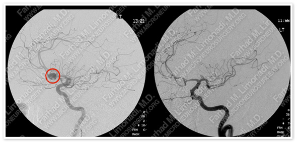
Post-op cerebral angiography shows complete treatment of this aneurysm.
Post-op Course
- She was discharged home and returned to normal function and her full time employment and remains free of any aneurysm.