


Case Presentation:
PCOM Aneurysm - Case 1
History and Physical
- 38-year-old lady who was found non-responsive and comatose in bed by her husband. She was intubated at the scene and brought to the hospital in an ambulance.
- On neurological examination, she was comatose and non-responsive.
Imaging
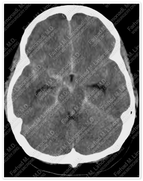
CT scan of the patient’s brain shows subarachnoid hemorrhage and hydrocephalus.
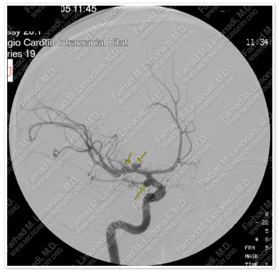
Cerebral angiography shows two right MCA (middle cerebral artery) and a PCOM (posterior communicating artery) aneurysms.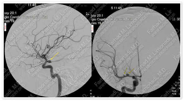
PCOM aneurysm is seen in the lateral projection on the left, and MCA aneurysms are seen on the right AP projection.
Surgical Procedure
- After placing a ventriculostomy in the ICU, she was taken to the operating room and underwent right orbitozygomatic craniotomy and micro-clipping of all three aneurysms.
Post-op Imaging
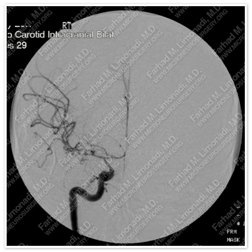
Post-operative cerebral angiography shows severe vasospasm (as the result of subarachnoid hemorrhage). This vasospasm was aggressively treated medically in the ICU with HHH therapy (Hypertensive, Hypervolemic, and Hemodilution). She responded to this treatment and gained consciousness.
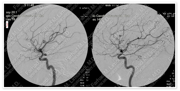
Follow-up cerebral angiography showed complete treatment of all the aneurysms and the vasospasm.
Post-op Course
- She was eventually weaned off the ventriculostomy and was discharged from the hospital. She fully recovered and remains aneurysm free and has returned to full time employment.