


Case Presentation:
PICA Aneurysm - Case 2
Ruptured Posterior Inferior Cerebellar Artery
History & Physical
- 61-year-old man presented with severe headache and somnolence.
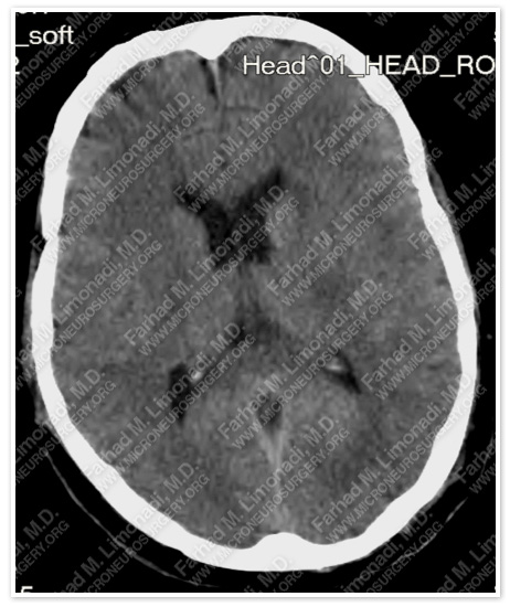
Imaging
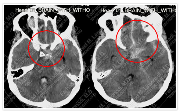
CT scan of the patient’s brain shows a subarachnoid hemorrhage.
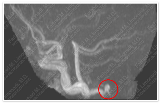
MRA image on the top and CTA image on the bottom show a left PICA (posterior inferior cerebrallar artery) aneurysm.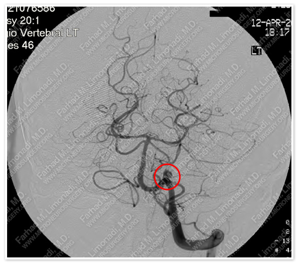
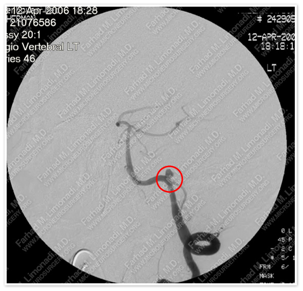
Cerebral angiography shows a PICA aneurysm. In addition, he lacks a right vertebral artery.
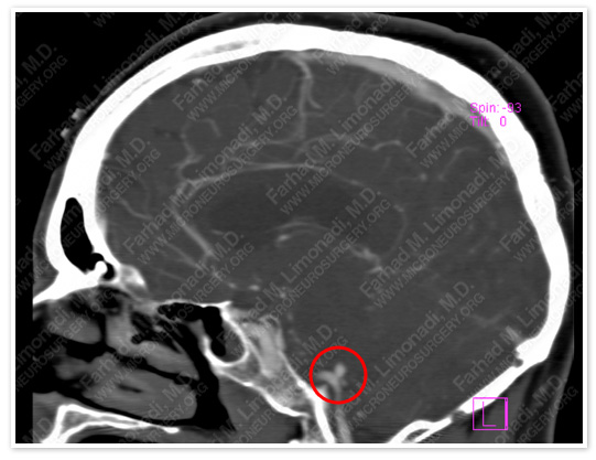
Computer Navigation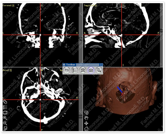
Computer navigation and stereotaxy was utilized to precisely model and map the aneurysm for surgical planning. The aneurysm is shown under the hairpin, and marked red in the right lower model of the skull.
Surgical Procedure
- He underwent left far lateral craniectomy with micro-clipping of this aneurysm utilizing intraoperative neurophysiological monitoring and stereotaxy.
Post-op Imaging
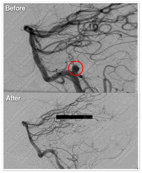
Post-op cerebral angiography shows complete treatment of this aneurysm.
Post-op Course
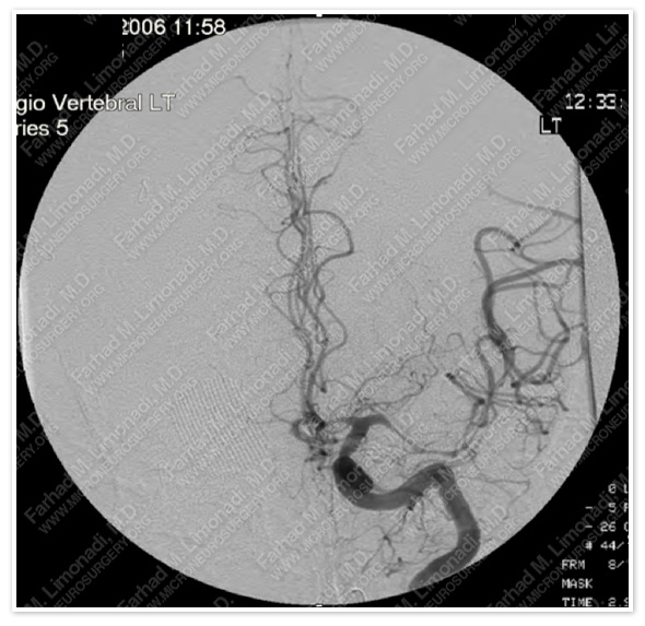
On post bleeding day #11, while being monitored in the ICU he developed weakness in his right side. Cerebral angiography showed severe vasospasm (as the result of subarachnoid hemorrhage).
On discharge, his CT showed clearance of subarachnoid hemorrhage.
- This vasospasm was aggressively treated medically in the ICU with HHH therapy (Hypertensive, Hypervolemic, and Hemodilution). He responded to this treatment and was discharged from the hospital.
- He has since returned to normal function and remains free of any aneurysm.