


Case Presentation:
Trigeminal Neuralgia & Microvascular Decompression - Case 1
History & Physical
- 69-year-old right-handed lady with initial onset of severe facial pain 20 years ago which resulted in multiple tooth extractions and eventually was correctly diagnosed as trigeminal neuralgia. She was experiencing burning and electricity type sensation with sensation of being struck on the face with an ice pick. The pain would be triggered by dental brushing, application of make-up, or at times chewing. She was appropriately managed with Tegretol, and following becoming refractory to this medication, Neurontin was also added to her medication regimen. However, her pain eventually returned despite this. She was diagnosed with typical trigeminal neuralgia.
- On examination, patient had no neurological deficit.
Imaging
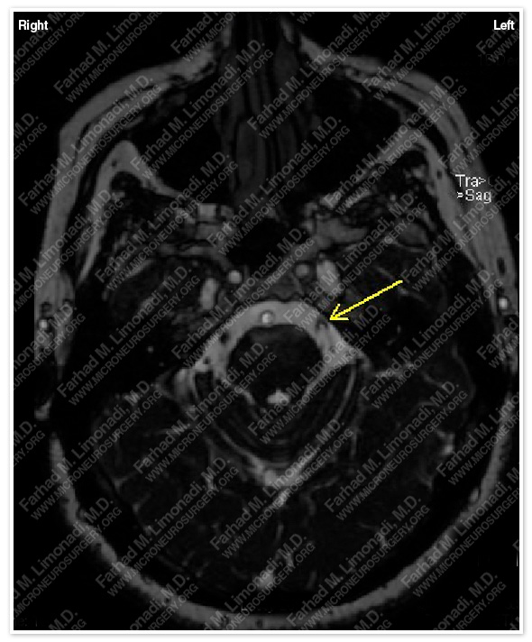
MRI scan of her brain demonstrated a vascular structure impinging and compressing the trigeminal nerve at root entry zone.
Surgical Procedure
- Having been correctly diagnosed with trigeminal neuralgia, and been refractory to proper medical management, she underwent microvascular decompression of the trigeminal nerve by left retrosigmoid craniectomy.
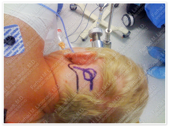
The planned bony exposure is marked in blue (circle), and important vascular and bony landmarks (mastoid tip, transverse and sigmoid sinuses) are also marked.
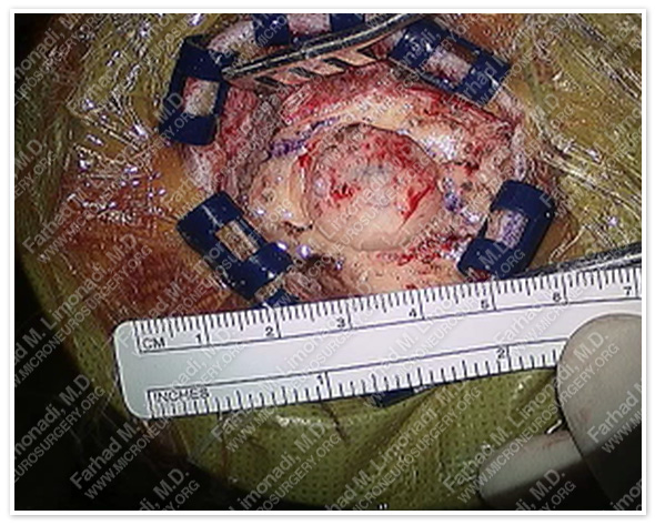
Retrosigmoid craniectomy is performed using micro-drill with bony exposure approximately the size of a quarter coin.
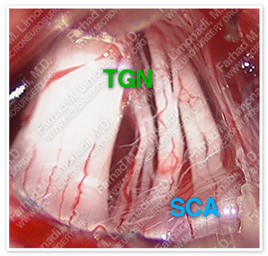
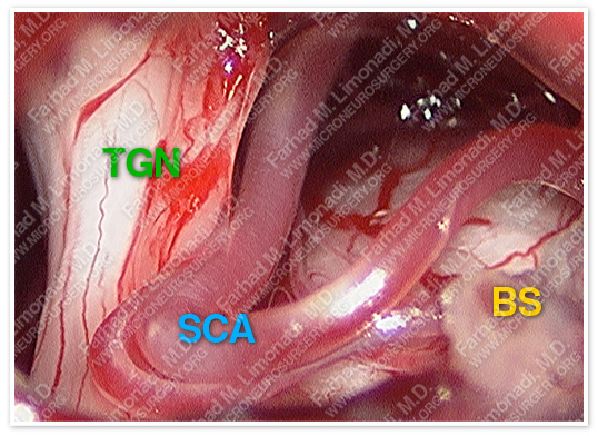
Trigeminal nerve (TGN) is distorted and compressed by a pulsating superior cerebellar artery (SCA).
Superior cerebellar artery (SCA) has been mobilized from its constrained position at root entry zone of trigeminal nerve (TGN) as the first step of microvascular decompression (MVD). Brain stem is seen in the bottom of the picture (BS).
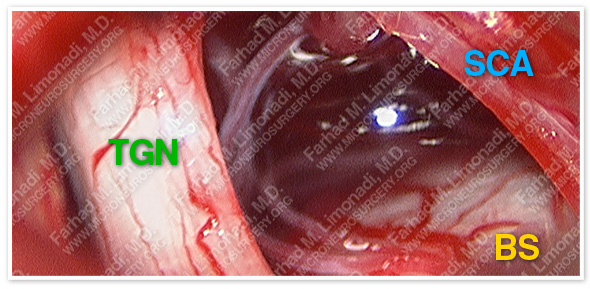
Trigeminal nerve (TGN) is completely separated from the pulsating superior cerebellar artery (SCA). Brain stem is seen in the bottom of the picture (BS).

Microvascular decompression (MVD) is completed by placing a teflon felt (TF) between the trigeminal nerve and the artery to ensure permanent separation of these structures.
Post-op Course
- Patient did well postoperatively with complete resolution of her facial pain. She was discharged from the hospital and returned to full function.