


Case Presentation: Trigeminal Neuralgia & Microvascular Decompression - Case 3
History and Physical
History and Physical
- 64 year old right handed lady with initial onset of sever facial pain 8 years ago which was appropriately managed with Tegretol initially. Subsequent to becoming refractory to this medication, Neurontin and Baclofen were also added to her medication regimen. However, her episodic pain persisted despite this. She was diagnosed with typical trigeminal neuralgia (or trigeminal neuralgia type 1).
- On examination patient had no focal neurological deficit.
Imaging
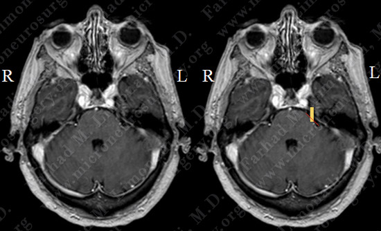
- MRI scan of her brain demonstrated a vascular structure (red) impinging and compressing the trigeminal nerve (yellow) at root entry zone.
Surgical Procedure
- Having been correctly diagnosed with trigeminal neuralgia, and been refractory to proper medical management , she underwent microvascular decompression of trigeminal nerve by left retrosigmoid craniectomy.
- This operation is performed by making a small opening (size of a quarter coin) in the skull and utilizing the surgical microscope to perform the surgery through this small opening.
- Intra-operative neurophysiological monitoring is used to monitor the function of 7th, and 8th cranial nerves.
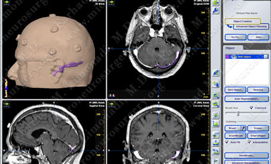
- Computer navigation and stereotaxy is also utilized
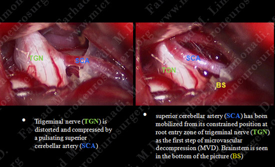
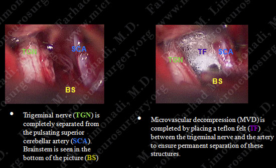
View through Surgical Microscope
Hospital Course
- Patient did well post-operatively with complete resolution of her facial pain. She was discharged from the hospital and returned to full function.