


Case Presentation:
Thoracic Schwannoma Spine Tumor - Case 1
History & Physical
- 59-year-old gentleman who presented to the emergency room with progressive loss of strength in his lower extremities and inability to walk over a short period of time. He was also was suffering from bowel and urinary incontinence with loss of sensation in his lower extremity and torso.
- On examination, he had significant weakness in both lower extremities, with numbness up to T7 level. He had no rectal tone.
Imaging
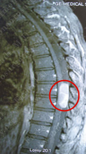
MRI scan of the patient’s thoracic spine shows a large intradural extramedullary tumor at T7 and T8 level compressing the spinal cord.
Surgical Procedure

He underwent T7, T8, and T9 laminectomy with surgical resection of this tumor using computer navigation, stereotaxy, and intraoperative neurophysiological monitoring.
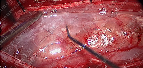
View of the tumor through the surgical microscope.
Pathology

The pathology of the tumor proved the diagnosis of spinal schwannoma.
Post-op Imaging
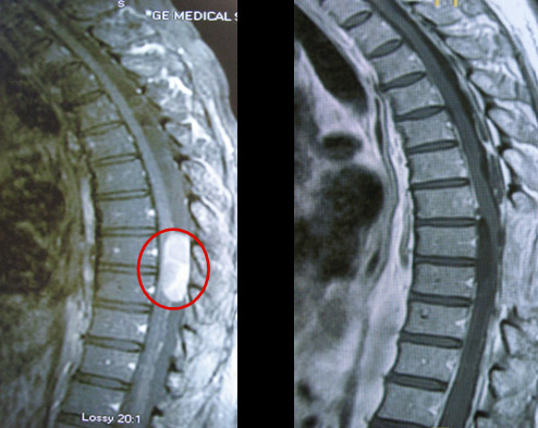
Before Operation After Operation
Post-op MRI shows complete resection of the tumor.
Post-op Course
- The patient did well postoperatively with regaining his ability to walk independently using a cane. His bowel and bladder function also returned to near normal function and his numbness was limited to his feet.