


Case Presentation:
ACOM Aneurysm - Case 3
Subarachnoid Hemorrhage
History & Physical
- 66-year-old left-handed lady who presented to the hospital with severe headache.
- On physical examination, she had no neurological deficit. She, however, had significant neck pain and meningismus.
Imaging
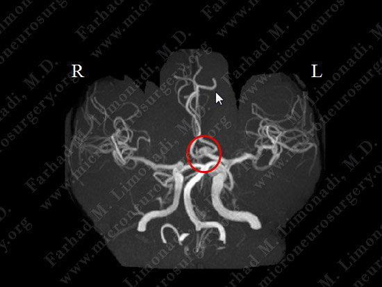
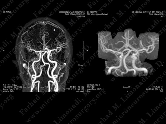
MRA (Magnetic Resonance Angiography) of the brain demonstrates an ACOM (anterior communicating) aneurysm (circled in red).
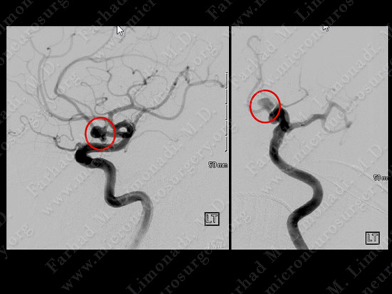
Cerebral angiography confirmed the diagnosis of ACOM aneurysm. This aneurysm is circled in red in lateral (left) and AP view (right).
3D cerebral angiography showing the anatomy of the aneurysm.
Computer Navigation
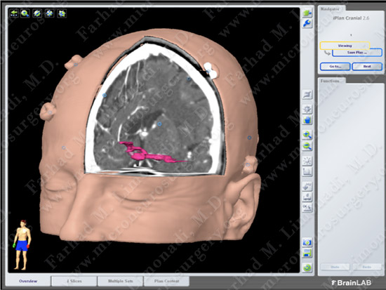
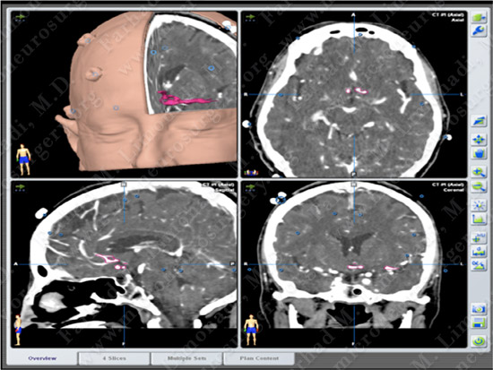
Computer simulation and modeling was utilized for precise localization of aneurysm and its vascular supply (depicted in pink).
Surgical Procedure
- A coil embolization of the aneurysm was attempted and failed due to unfavorable aspect ratio of the aneurysm. Therefore, she instead underwent right supraorbital skull base supraorbital craniotomy with micro-clipping of the aneurysm in the operating room utilizing intra-operative neurophysiological monitoring and brain mapping.
Supraorbital craniotomy.
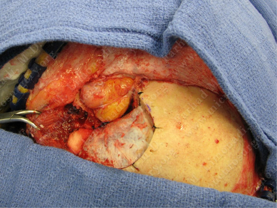
Exposure of the dura after removal of the orbital roof and wall.
Dura opened and orbital content is advanced anteriorly.
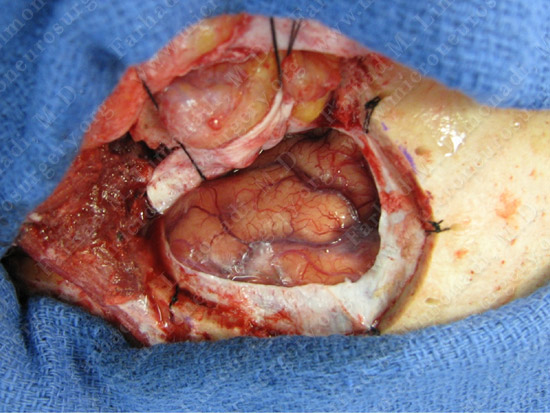
View through the microscope; micro-clipping of the aneurysm.
Post-op Imaging
Post-op MRA shows complete treatment of the aneurysm with no evidence of filling.
Post-op Course
- Patient returned to complete normal function and was discharged home