


Case Presentation:
Cervical Stenosis - Case 2
History & Physical
- 73-year-old right-handed man who presented with inabliity to walk, feed or groom himself.
- His neurological examination showed significant weakness of upper extremities with atrophy of dorsal interosseous muscles. He was non-ambulatory.
Imaging
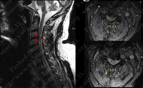
MRI scan of the patient's cervical spine (neck) shows severe stenosis (narrowing) at C3-C4 with evidence of spinal cord compression and contusion.
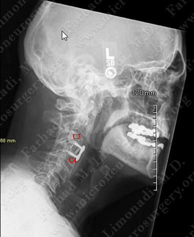
Surgical Procedure
- After a comprehensive work-up and eliminating other potential contributing causes of his disease, and an appropriate period of conservative measures, he underwent anterior cervical discectomy and instrumented fusion (ACDF) at C34, C45 and C56.
Post-op Course
- He did well postoperatively with immediate improvement of his function. He was able to walk, feed and groom himself.