


Case Presentation:
Choroid Plexus Papilloma - Case 1
- 35-year-old lady who presented with sever headache and photophobia.
- On examination she had no focal neurological deficit.
- MRI scan of her brain was obtained which showed a well circumscribed contrast enhancing tumor in the fourth ventricle.
Imaging
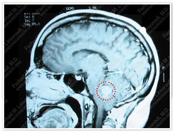
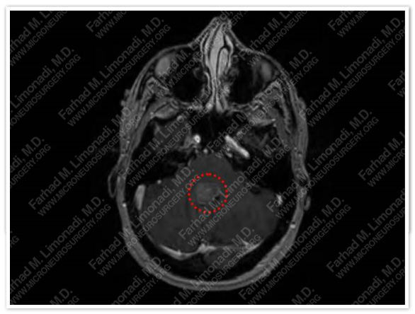
MRI of the brain showed a well circumscribed contrast enhancing tumor in the fourth ventricle.
The tumor is outlined with a dotted red circle.
Computer Navigation
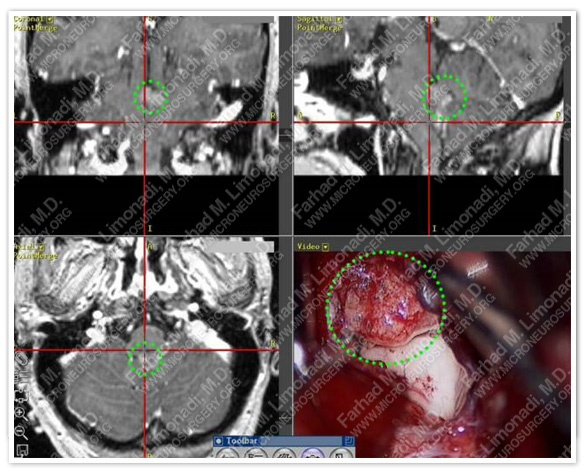
Stereotactic Image guidance was used to identify and resect the tumor. The tumor is being resected off the brainstem and is shown in a dotted green circle.
Surgical Procedure
- She underwent suboccipital craniectomy and Telovelar approach to the fourth ventricle with gross total resection of the tumor
- This operation was performed using:
1 - Stereotactic image guidance
2 - Intraoperative neurophysiological monitoring of lower cranial nerves (9, 10, 11, and 12) and BAER (Brain Stem Auditory Evoked Response).
3 - Neuroendoscopy
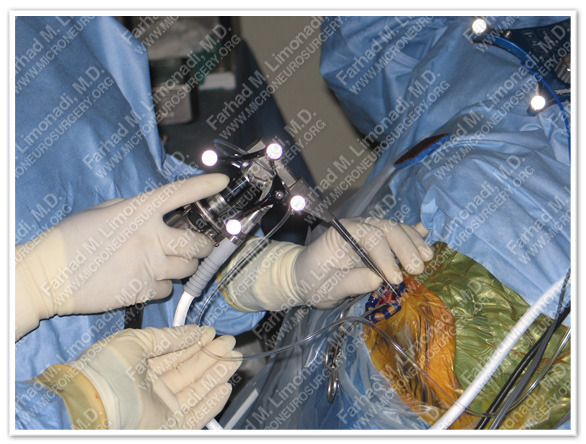
Stereotactic Neuroendoscopy is used to see and resect tumor from hard to see corners in the 4th ventricle.
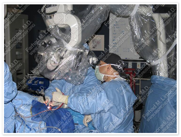
Patient is placed in prone position and using surgical microscope suboccipital craniectomy following exposure of cerebellar tonsills are performed.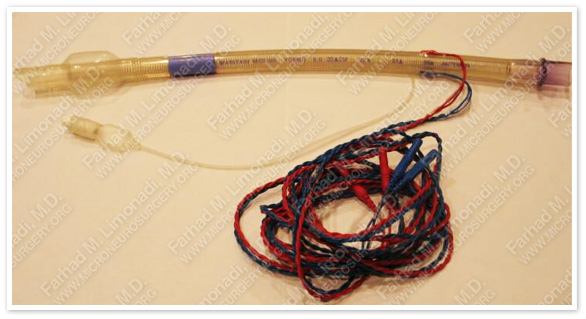
A special tracheal tube with electrical circuit is utilized for monitoring of cranial nerves 9 and 10.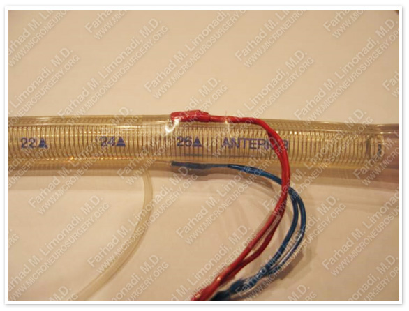
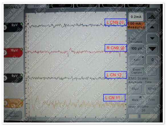
Cranial nerves 9, 10, 11 and 12 are monitored carelfully while dissecting the tumor off the brainstem.
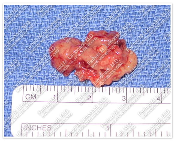
The slide above shows the tumor which was removed in gross total fashion and in one piece.
Pathology
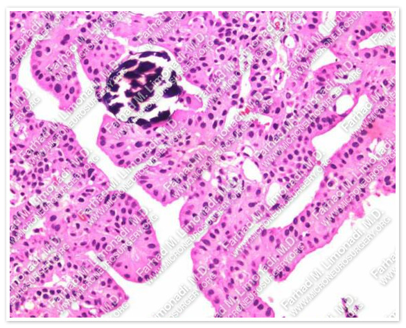
The slide above shows the H&E stain of the tumor. The cauliflower papillary shape with cuboidal and columnar cells is typical features of choroid plexus papilloma.
Post-op Imaging
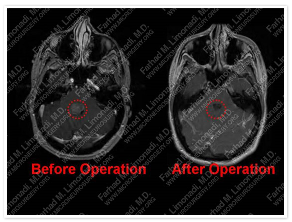
Axial MRI of the brain before and after shows complete resection of tumor.
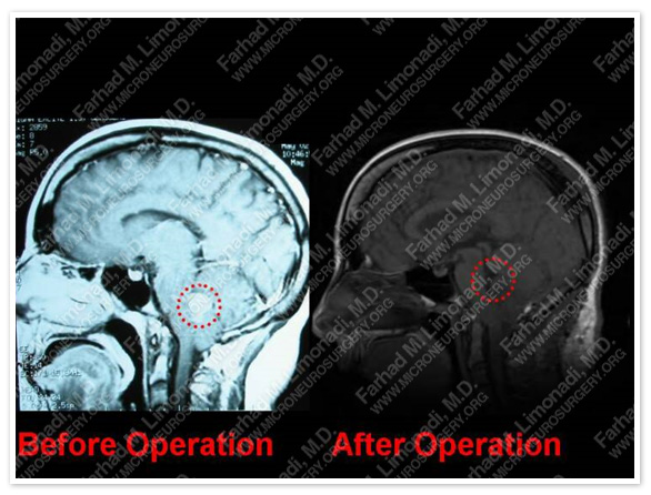
Sagittal MRI of the brain before and after shows complete resection of tumor.
Post-op Course
- Patient did well postoperatively with mild double vision which improved shortly thereafter.