


Case Presentation:
Glioblastoma Multiforme (GBM) - Case 1
History & Physical
- 55-year-old lady presented with new onset seizure.
Imaging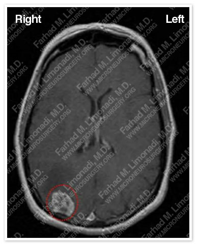
An MRI of her brain disclosed a right occipital tumor (outlined in a red dotted circle).
Surgical Procedure
- She underwent frameless stereotactic craniotomy with gross total resection of the tumor which was later found to be glioblastoma multiforme (GBM).
Computer Navigation
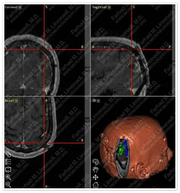
Computer navigation shows a virtual window through the skull over the tumor. The actual craniotomy size was as small as the tumor.
Post-op Imaging
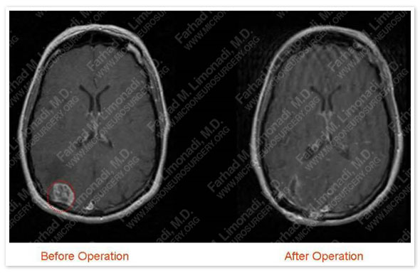
Postoperative MRI showed complete resection of the radiographically apparent tumor. Tumor is outlined in a red dotted circle.
Post-op Course
- She was discharged home three days later in good health and with no postsurgical complications.