


Case Presentation:
Hemangioblastoma - Case 1
History & Physical
Hemangioblastoma - Case 1
History & Physical
- 77-year-old man who presented with frequent falling and dizziness.
Imaging
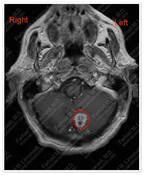
MRI scan of his brain showed a cystic mass in the left cerebellum.The tumor is outlined in a red dotted circle.
Computer Navigation
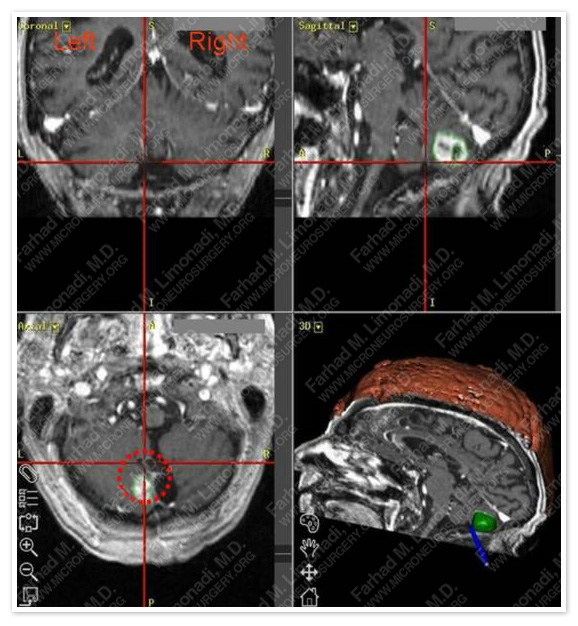
Tumor is volumetrically mapped and shown in green by 3D reconstruction of patient’s head in the right lower picture during the operation.
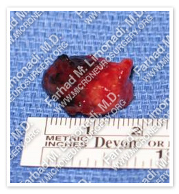
Surgical Procedure
- He underwent computer assisted frameless stereotactic craniectomy with complete resection of the tumor.
Post-op Imaging
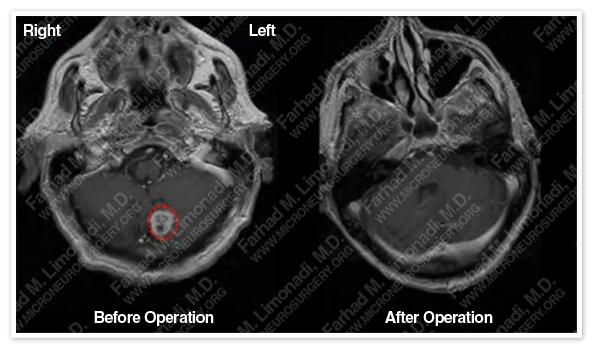
Post-op MRI showed complete resection of the tumor.
Post-op Course
- Patient was discharged in a few days in good health and with no postsurgical complications. His symptoms caused by the tumor were completely resolved and he returned to normal function with no dizziness or difficulty walking.