


Case Presentation:
Glioblastoma Multiforme (GBM) - Awake Craniotomy - Case 11
History and Physical
- 71 year old lady who presented with first onset of tonic-clonic seizure.
- On physical examination she had no focal neurological deficit.
Imaging
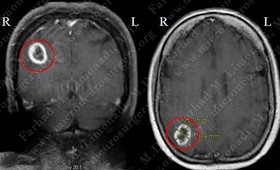
MRI scan of patient's brain shows a right parietal ring enhancing tumor (within the red circle) with significant vasogenic edema and local mass effect.
Surgical Procedure
- She underwent surgical resection of this tumor utilizing brain mapping, stereotactic and computer navigation, and intra-operative neurophysiological monitoring.
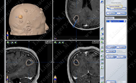
Stereotactic and computer navigation was used to determine the precise location of the tumor (outlined in yellow).
Pathology
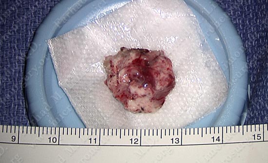
Tumor was removed in a gross total fashion.
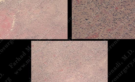
The pathology of the tumor confirmed diagnosis of GBM.
Post-op Imaging
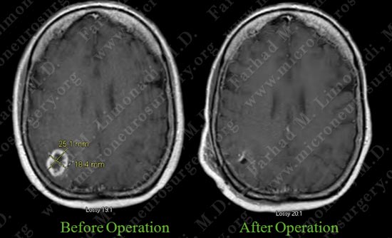
Post op MRI shows complete resection of the tumor with no injury to surrounding neuro-vascular structures.
Post-op Course
- Post operatively patient returned to completely normal neurological status with no complications. She was discharged home and returned to work.