


Case Presentation:
Glioblastoma Multiforme (GBM) - Case 12
History and Physical
- 60+ year-old lady who presented with new onset of tonic-clonic seizure.
- On physical examination, she has no focal neurological deficit with exception of diminished vision in the left inferior visual field (left inferior quadranopsia).
Imaging
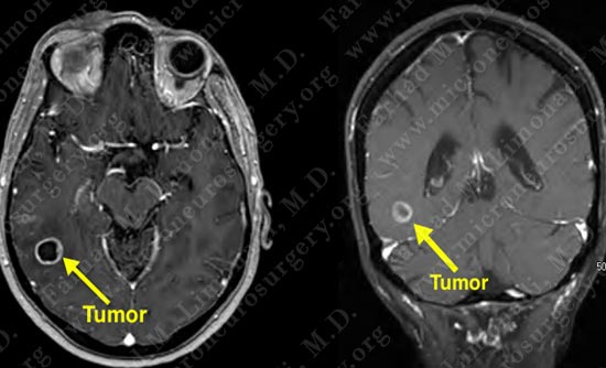
MRI scan of her brain shows a right posterior temporal contrast enhancing mass (tumor). The tumor is marked in the left side of the slides (right side of patient's brain).
Treatment Plan
- Patient was indicated for surgical resection of the tumor.
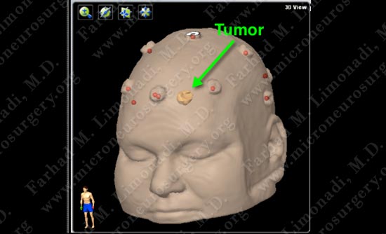
- Given the location of the tumor the plan was made to proceed with a posterior temporal approach utilizing computer navigation, stereotaxy, and intra-operative electrophysiological monitoring.
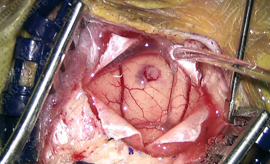
Surgical Procedure (view through the surgical microscope)
- She underwent right temporal craniotomy. Computer navigation was utilized to elevate a small bone flap of approximately 1 inch in diameter.
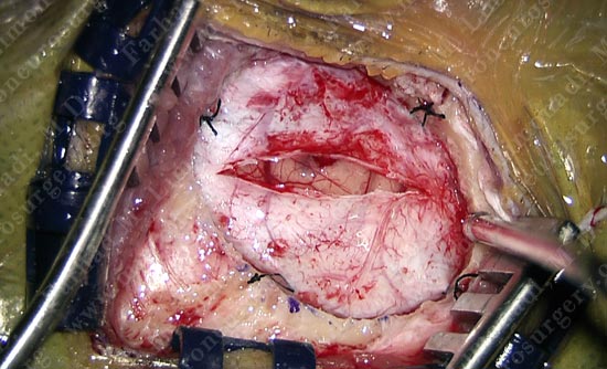
Dura is opened with temporal brain brought to view through a one inch window.
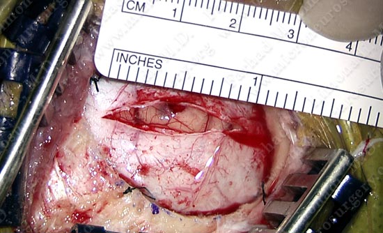
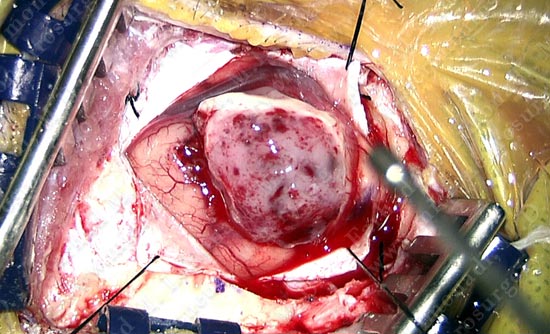
Tumor is removed from the right temporal lobe. It is almost as large as the surgical window.
Pathology
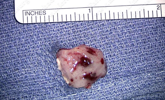
Tumor was removed in a gross total fashion.
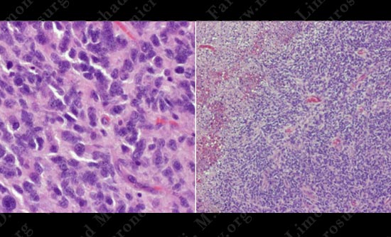
Pathology of the tumor was that of a Glioblastoma Multiforme (GBM). Given the pathology, additional tissue was removed from the cavity to have a tumor free margin.
Post-op Imaging
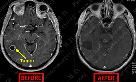
Post operative MRI shows complete resection of tumor with appropriate margin.
Post-op Course
- Patient did well post-operatively with no new neurological deficit. She was discharged.
- In four weeks she underwent radiotherapy and chemotherapy for adjuvant treatment of the tumor.