


Case Presentation:
Glioblastoma Multiforme (GBM) - Case 3
History & Physical
- 64-year-old right-handed gentleman presented to the hospital with the inability to speak.
- On examination, he had expressive and receptive aphasia (mute), with weakness of the right side, along with memory and cognitive decline.
Imaging
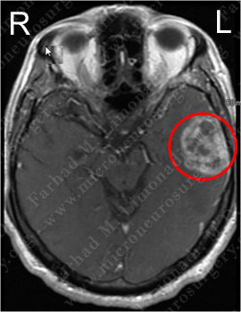
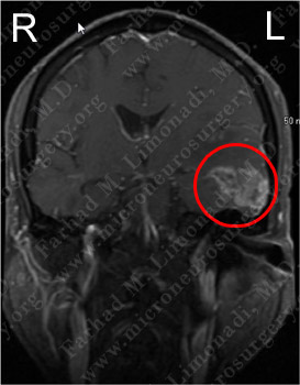
MRI scan of the patient's brain showed a left temporal brain tumor.
Treatment
- His speech improved with administration of steroids before the operation.
Surgical Procedure
- He underwent awake surgical resection of this tumor utilizing brain mapping, cortical stimulation, stereotatic and computer navigation, and intraoperative neurophysiological monitoring.
Computer Navigation
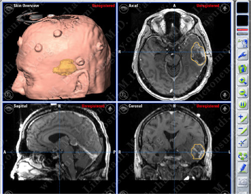
Computer navigation and stereotaxy utilized to map and localize the tumor (outlined in yellow) during surgery.
Pathology
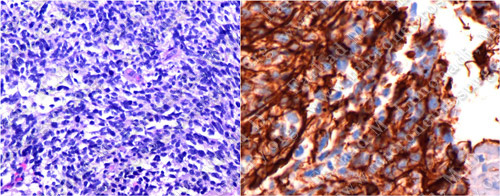
The pathology of the tumor confirmed diagnosis of Glioblastoma Multiforme (GBM).
Post-Op Imaging
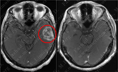
Post-op MRI shows complete resection of the tumor with no injury to surrounding neurovascular structures.
Post-op Course
- The patient did well postoperatively with no neurological deficit. His speech returned nearly to his baseline.