


Case Presentation:
Glioblastoma Multiforme (GBM) - Case 4
- 77-year-old gentleman with a new onset of tonic-clonic seizure. He was brought to the emergency room and after seizure control an MRI scan of his brain was performed.
- On examination he had right hemiparesis (weakness), however, was otherwise neurologically intact.
Imaging
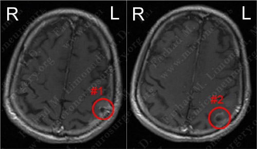
MRI scan of the patient's brain showed two contrast-enhancing tumors in close proximity to each other.
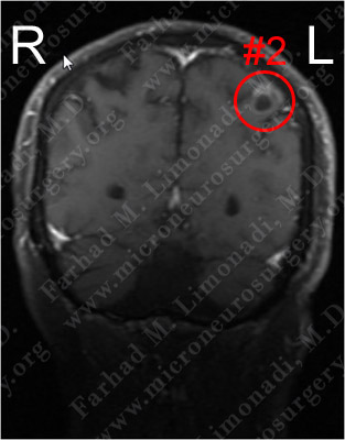
Coronal view shows the second lesion to a better advantage.
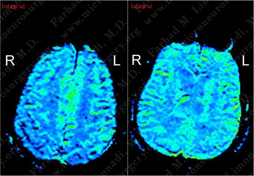
CT SPECT does not show significant hypermetabolism of either of the lesions.
Computer Navigation
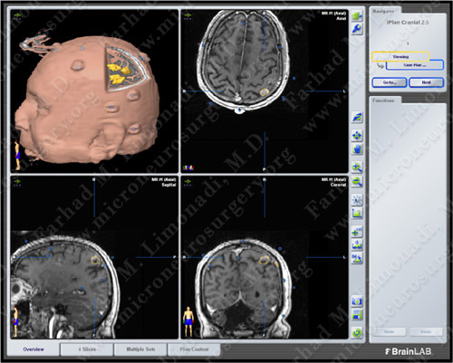
Computer navigation and stereotaxy utilized to map and localize the tumors (outlined in yellow) during surgery.
Surgical Procedure
- He underwent surgical resection of these tumors utilizing brain mapping, stereotactic and computer navigation, and intraoperative neurophysiological monitoring.
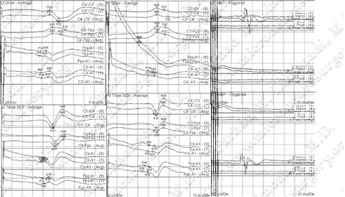
Intraoperative neurophysiological monitoring was utilized to maintain the integrity of his neurological functions
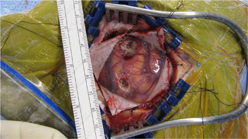
Utilizing computer navigation, a small craniotomy was performed precisely over both tumors and both tumors were removed using this small opening.
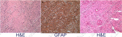
The pathology of the tumor showed necrosis (left), positive GFAP stain (center), and vascular proliferation (right), all of which revealed the diagnosis of Glioblastoma Multiforme (GBM).
Post-Op Imaging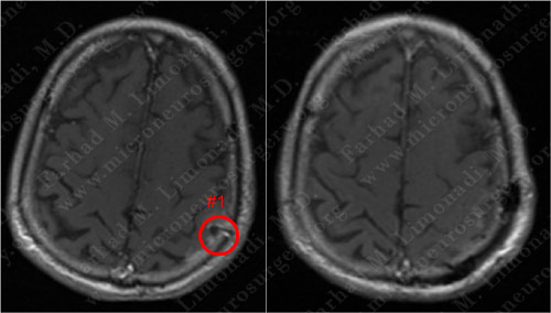
Before Operation Afer Operation
Post-op MRI shows radiographic resection of the first tumor.
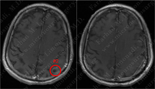
Before Operation After Operation
Post-op MRI shows radiographic resection of the second tumor as well.
Post-op course
- The patient did well postoperatively and was discharged home with steroids and anticonvulsants. His weakness completely resolved. However, due to the malignant nature of this tumor, he underwent conformal radiotherapy of the regions of both tumors.