


Case Presentation:
Glioblastoma Multiforme (GBM) - Case 5
History & Physical
- 50-year-old gentleman presented to the emergency room with loss of comprehension and ability to speak (global aphasia).
- On examination, he had global aphasia.
Imaging
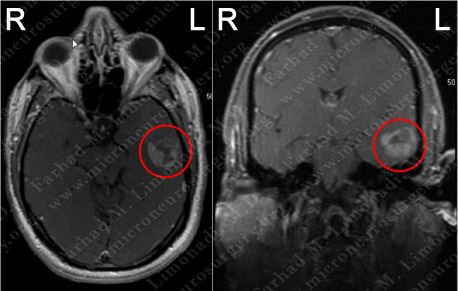
MRI scan of the patient's brain showed a large left temporal brain tumor.
Computer Navigation
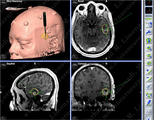
Computer navigation and stereotaxy utilized to map and localize the tumors (outlined in yellow) during surgery.
Surgical Procedure
- He underwent a left temporal craniotomy and surgical resection of the tumor utilizing brain mapping, stereotactic and computer navigation, and intraoperative neurophysiological monitoring.
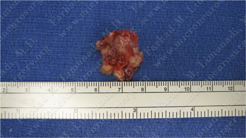
Tumor specimen
Pathology
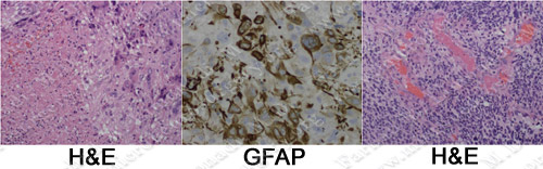
The pathology of the tumor showed necrosis (left), positive GFAP stain (center), and vascular proliferation (right) all of which revealed the diagnosis of glioblastoma multiforme (GBM).
Post-op Imaging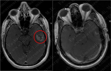
Before Operation After Operation
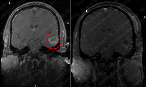
Before Operation After Operation
Post-op MRI shows radiographic resection of the tumor.
Post-op course
- The patient did well postoperatively and was discharged home with steroids and anticonvulsants. His speech returned to normal. However, due to the malignant nature of this tumor, he underwent conformal radiotherapy of the cavity at the tumor resection site.