


Case Presentation:
Glioblastoma Multiforme (GBM) - Case 8
History & Physical
-
77-year-old right-handed gentleman who is stauspost-resection of GBM in left frontoparietal brain five months ago. He did well post-operatively and subsequently underwent conformal radiotherapy. He presented with seizure refractory to conservative measures and despite two antiepilepctic drugs.
-
On examination, he had no focal neurological deficit.
Imaging
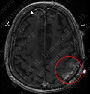
MRI scan of the patient’s brain showed two nodular enhancements near motor cortex diagnosed as recurrent tumors.
Computer Navigation
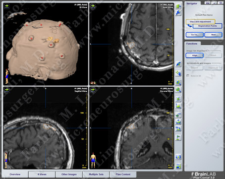
Computer navigation and stereotaxy utilized to map and localize the tumor (outlined in yellow) during surgery.
Surgical Procedure
-
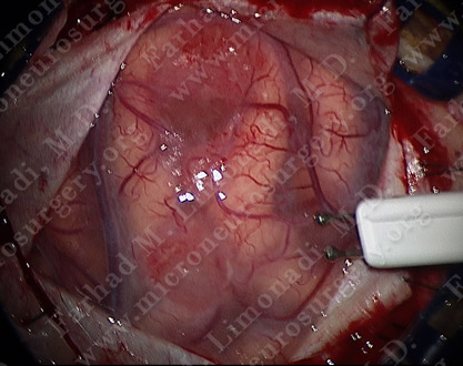
He underwent surgical resection of both of these nodular enhancing tumors utilizing brain mapping, cortical stimulation, stereotactic and computer navigation, and intraoperative neurophysiological monitoring.
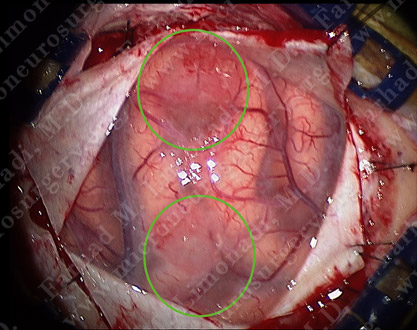
View through the surgical microscope shows two nodular tumors (outlined by green circles).
Motor cortex stimulation using Ojeman stimulator.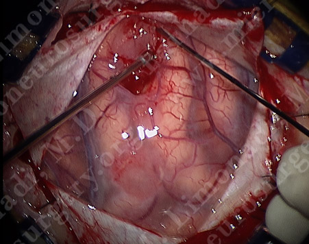
One of the tumors being removed.
Utilizing computer navigation, a small craniotomy was performed precisely over the tumor and both tumors were removed using this small opening.
Pathology
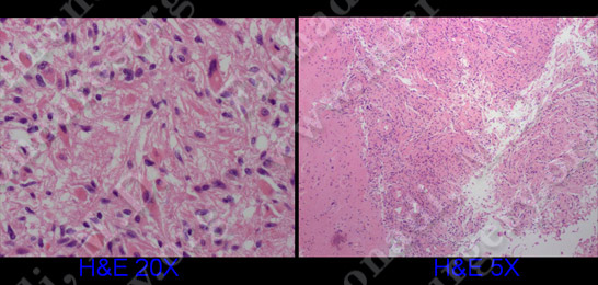
The pathology of the tumor confirmed diagnosis of glioblastoma multiforme (GBM).
Post-op Imaging
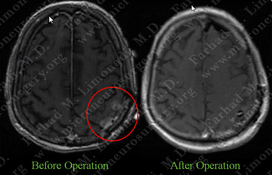
Post-op MRI shows complete resection of both tumors with no injury to surrounding neurovascular structures.
Post-op Course
-
The patient did well postoperatively with no neurological deficit. He was discharged home.