


Case Presentation:
Medulloblastoma Brain Tumor - Case 1
History & Physical
- 20+ year old man presented with headache, nausea and vomitting.
- On examination, he had no focal neurological deficit and was completely alert and oriented.
Imaging
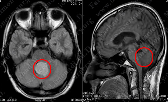
MRI scan of the patient’s brain shows a tumor within the fourth ventricle impinging on the brain stem and mild hydrocephalus.
Computer Navigation
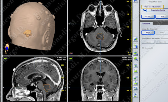
Stereotaxy and computer navigation was utilized to localize and remove the tumor precisely, with surrounding neurovascular structures mapped out and protected.
Surgical Procedure
- Sub-occipital inter-tonsillar approach was utilized.
Pathology
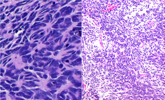
The pathology of the tumor confirmed the diagnosis of medulloblastoma.
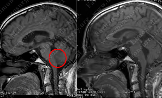
Post-op Imaging
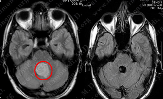
Before Operation After Operation
Before Operation After Operation
Post-op MRI shows complete resection of the tumor with no injury to surrounding neurovascular structures and resolution of hydrocephalus.
Post-op Course
- The patient did well postoperatively with no neurological deficit. He was discharged home and returned to full employment.