


Case Presentation:
Oligodendroglioma - Case 1
History & Physical
- 32-year-old lady with known diagnosis of oligodendroglioma in right occipitoparietal region and three previous subtotal resections of this tumor who was presented to us for complete resection.
Imaging
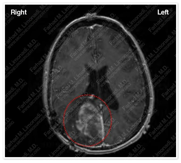
The tumor is outlined in a red dotted circle.
Surgical Procedure
- She underwent frameless stereotactic craniotomy and subtotal resection of the tumor.
- Virtual imaging determined the precise location of the tumor and important adjacent structures.
- The tumor was resected via a small occipital craniotomy.
Computer Navigation
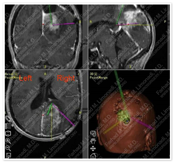
Volumetric map of the tumor assisted in safe and subtotal resection of the tumor while safeguarding vital neurovascular structures.
Pathology
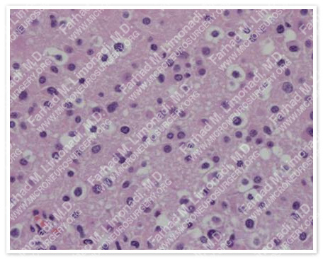
Histopathological study of the tumor confirmed previous diagnosis of oligodendroglioma.
Post-op Imaging
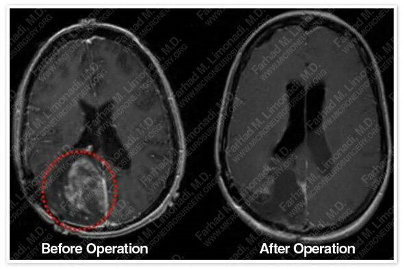
Postoperative MRI showed complete resection of the tumor.
Post-Op Course
- She was discharged home in three days in good health and with no postsurgical complication.