


Case Presentation:
Colloid Cyst - Case 1
History & Physical
- 60-year-old man who presented with nausea and vomiting for several days and decreased level of consciousness. CT scan of his brain showed severe hydrocephalus with dilated ventricles.
- He emergently underwent R frontal ventriculostomy as a temporizing measure to avoid brain herniation and death. The tip of the ventricular catheter is outlined in red.
Imaging
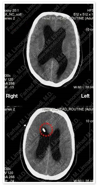
CT scan of his brain showed severe hydrocephalus with dilated ventricles. The ti pof the ventricular catheter is outlined in red.
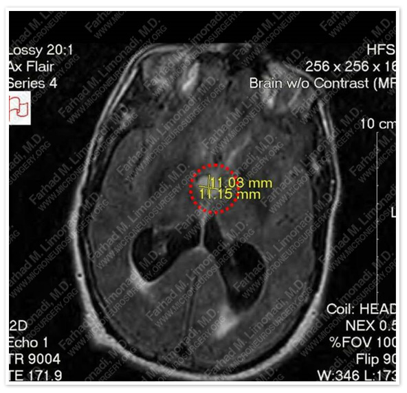
MRI of the brain showed a cystic lesion obstructing the foramen of Monro, and thereby, causing hydrocephalus. The colloid cyst is outlined in a red dotted circle.
Surgical Procedure
- He underwent frameless stereotactic transcallosal craniotomy with complete resection of the colloid cyst.
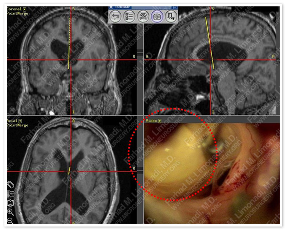
The colloid cyst is outlined in a red dotted circle in the view through the surgical microscope as it is being resected.
Post-op Imaging
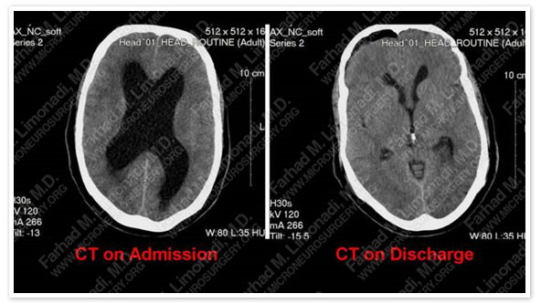
CT of the brain on discharge showed complete resolution of hydrocephalus.
Post-op Course
- Patient was discharged home in a few days in good health and with no postsurgical complication and complete resolution of his symptoms.