


Case Presentation:
Pituitary Tumors - Case 2
Macroadenoma
History & Physical
-
37-year-old man with progressive loss of vision resulting in becoming disabled. He lost his ability to read and write as the result and could no longer drive. He was admitted to the hospital through the emergency room.
-
On physical examination, he had a significant loss of vision acuity and had bi-temporal hemianopsia (blind in lateral visual field). He also had right sixth nerve palsy.
Imaging
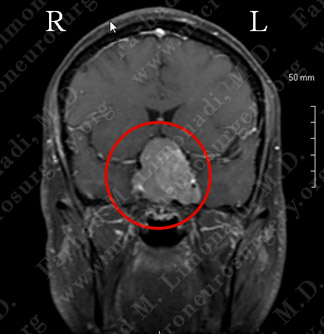
MRI scan of patient’s brain shows a large pituitary tumor with significant pressure on optic chiasm and mass effect on carotid arteries.
Computer Navigation
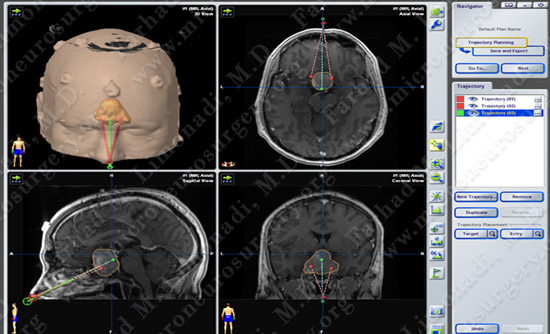
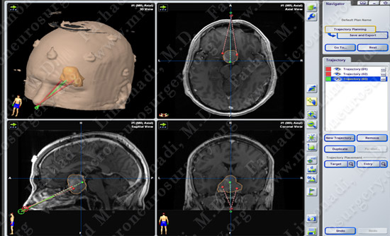
Computer simulation and modeling was utilized for precise localization of tumor (green in the right lower pic, and outlined in red circle).
Surgical Procedure
-
He underwent orbitozygomatic craniotomy and surgical resection of this brain tumor using brain mapping, stereotactic and computer navigation, and intraoperative neurophysiological monitoring.
Post-op Imaging
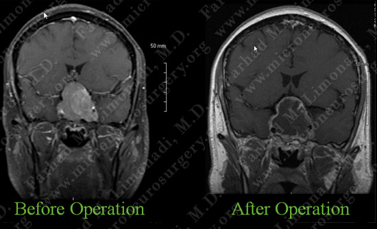
Post-op MRI shows complete resection of the tumor with no injury to surrounding neurovascular structures.
Post-op Course
- The patient did well and was discharged from the hospital with no complications. His vision has returned to near normal level and is regaining his ability to read and write.