


Case Presentation:
Pituitary Tumors - Case 3
Pituitary Apoplexy
History & Physical
-
The patient is a 31-year-old right-handed gentleman who noted severe headache as well as blurry vision approximately three weeks ago. He saw an ophthalmologist who discovered bi-temporal visual field cut and was referred for an MRI scan of his brain. His MRI showed a pituitary tumor with hemorrhage (pituitary apoplexy). The patient states that his vision has been blurry. He was complaining of progressive loss of vision and severe headache.
-
On examination he had bi-temporal visual field loss.
Imaging
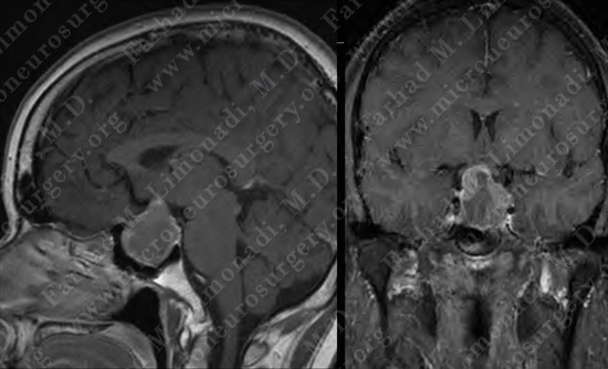
MRI scan of the patient’s brain shows a pituitary tumor with intratumoral hemorrhage and necrosis, otherwise known as pituitary apoplexy.
Computer Navigation
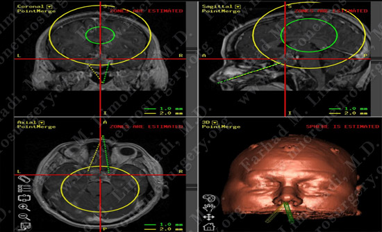
Computer simulation and modeling was utilized for precise localization of tumor with optimal approach.
Pathology

Pathology of the tumor was that of a benign prolactin-secreting pituitary adenoma.
Post-op Imaging
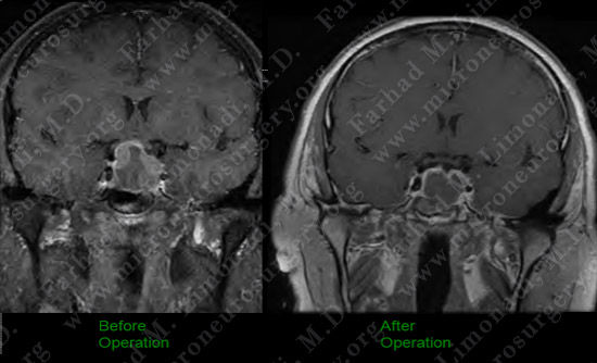
Post-op MRI shows complete resection of the tumor with no injury to surrounding neurovascular structures.
Post-op Course

Patient did well postoperatively with complete resolution of his symptoms. He returned to normal function and remained tumor free.