


Case Presentation:
Acoustic Neuroma - Case 3
- 70-year-old right-handed lady who was experiencing progressive difficulty with gait and balance for a month. This resulted in her requiring the use of a walker for ambulation. Further, she experienced a progressive loss of hearing in the right ear for three months. The patient had been experiencing urinary incontinence for a month as well as progressive loss of memory and difficulty with cognition and cognitive status. She came to the emergency room with eventual inability to ambulate and declining cognitive status.
- On physical examination, she had right facial weakness, hearing loss, and her gait was wide-based and unsteady.
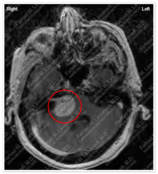
MRI scan of patient’s brain shows a large right acoustic neuroma with impingement of brain stem. She also has hydrocephalus.
- She underwent a right translabyrinthine craniectomy and surgical resection of this brain tumor using stereotaxy and brain mapping, intraoperative neurophysiological monitoring including facial nerve monitoring, SSEP, MEP, and brain stem auditory evoked response (BAER).
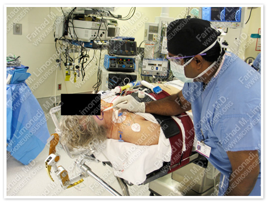
Patient is being prepared for surgery.
Computer Navigation
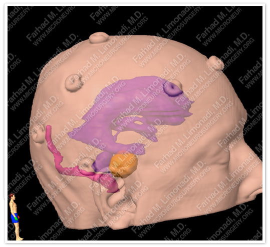
Computer navigation is used to study the precise location of the tumor with regard to important adjacent neurovascular structures. The tumor is colored yellow in this picture.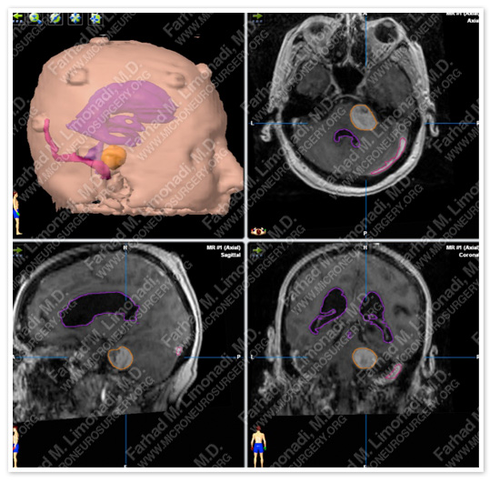
The computer model is fused with patient’s MRI for precise localization of tumor. The tumor is outlined in yellow in the MRIs.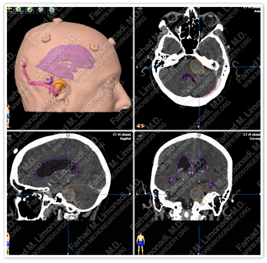
The computer model is fused with patient’s CT for precise localization of the tumor and study of patient’s bony anatomy. The tumor is outlined in yellow in the MRIs.
Pathology
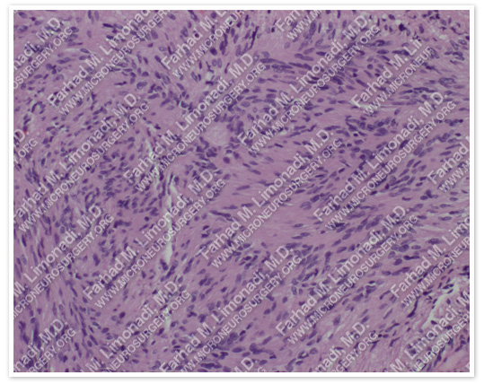
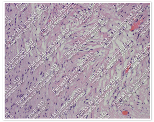
The pathology of the tumor confirmed diagnosis of acoustic neuroma (vestibular schwannoma).
Post-op Imaging
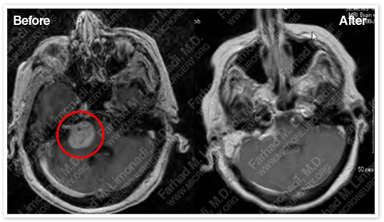
Post-op MRI confirms subtotal resection of the tumor with no injury to surrounding neurovascular structures.
Post-op Course
- Postoperatively, the patient’s cognitive status completely returned to normal level. She was able to ambulate independently and at times used a walker for support only. Her facial weakness nearly completely returned to normal with only a mild facial grimace asymmetry.