


Case Presentation:
Acoustic Neuroma - Case 7
History & Physical
- 33-year-old gentleman who presented with transient expressive aphasia versus dysarthria (inability to speak) upto four times a day and each time lasting 30 seconds. He had difficulty with rolling his tongue during these episodes which were observed by his colleagues and wife.
- On physical examination, he had no focal neurological deficit.
Imaging
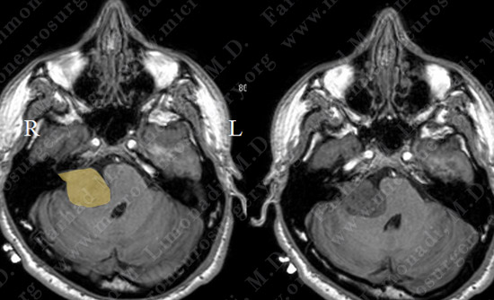
MRI scan of the patient’s brain shows a large vestibular schwannoma (acoustic neuroma) measuring 3 cm in the largest dimension (outlined in yellow). The tumor is deforming the brainstem and causing effacement of fourth ventricle.
Treatment
- He initially underwent treatment with steroids to help with edema and with the knowledge that this pattern of radiographic change and edema typically is followed by tumor shrinking in size.
- However, after nearly a month of steroid administration and continued surveillance, patient’s symptoms worsened and a follow-up MRI showed progressive enlargement of the tumor and mass effect on the brain stem.
Surgical Procedure
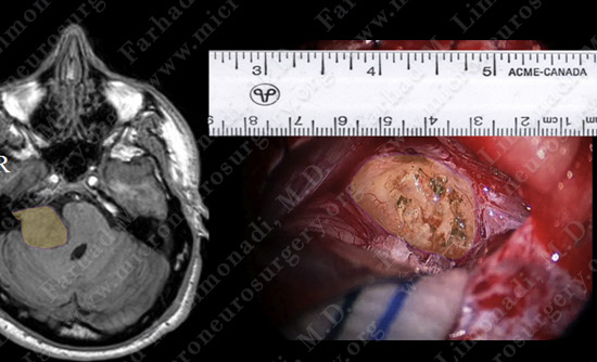
MRI showing the tumor (colored in yellow). View of the tumor through the microscope during surgery.
Computer Navigation
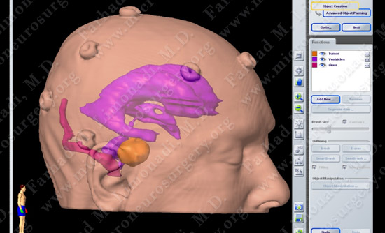
Computer navigation utilized during the surgery shows the tumor (yellow), vascular sinuses (red) and the ventricles (purple).
Post-op Course
- Postoperatively, the patient initially had no facial weakness. However, due to inflammation of the facial nerve, he did develop facial weakness with maintaining his ability to close his right eye. He no longer experienced episodes of dysarthria and speech difficulties. He was discharged home and has returned to full time-employment. It is expected that with time he will regain most of his facial nerve function.