


Case Presentation:
Acoustic Neuroma - Case 4
History & Physical
- 54-year-old lady presented to the emergency room with headache, dizziness, right facial weakness, and progressive difficulty with swallowing.
- On physical examination, she had House-Brackmann Grade IV facial weakness on the right as well as no hearing on the right side.
Imaging
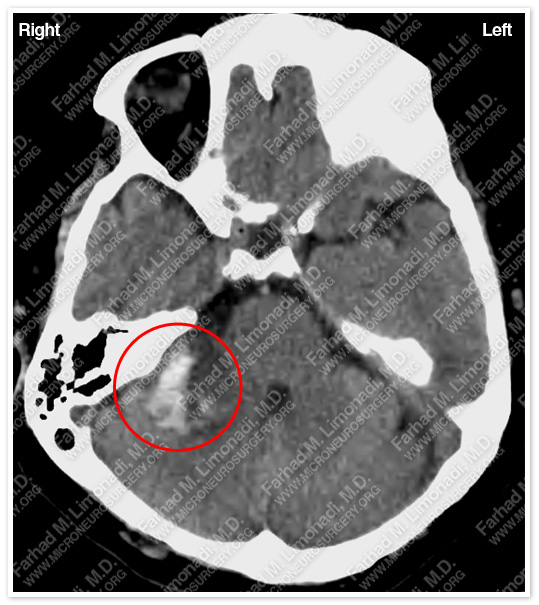
CT scan of the patient’s brain shows a right hemorrhagic cerbellopontine angle tumor pressing on the brain stem.
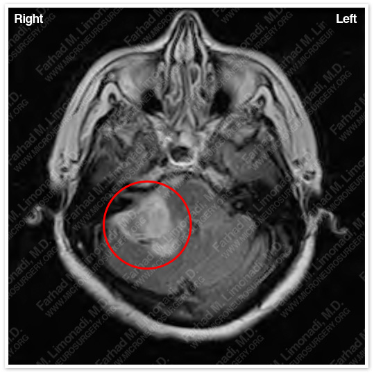
MRI scan of the patient’s brain shows a large right acoustic neuroma with central necrosis and hemorrhage.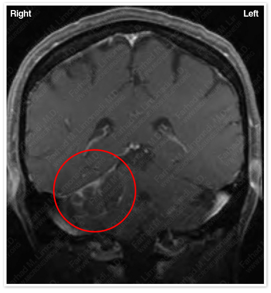
MRI scan flair sequence shows significant vasogenic edema involving brain stem and cerebellum.
Surgical Procedure
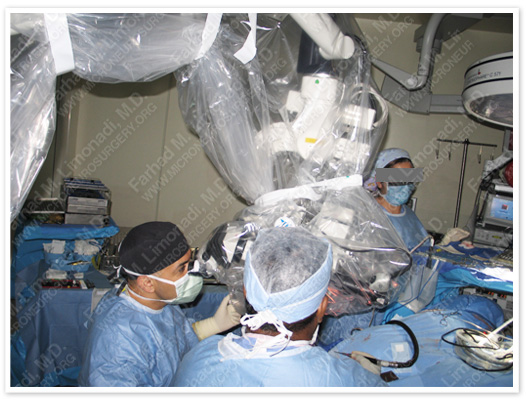
- She underwent a right retrosigmoid craniectomy and surgical resection of this brain tumor using intraoperative neurophysiological monitoring including facial nerve monitoring and brain stem auditory evoked response (BAER).
Pathology
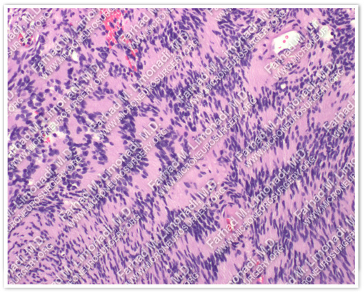
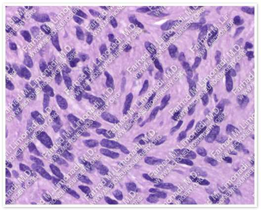
The pathology of the tumor confirmed diagnosis of acoustic neuroma (vestibular schwannoma).
Post-op Imaging
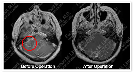
Post-op MRI confirms complete resection of the tumor with no injury to surrounding neurovascular structures.
Post-op course
- The patient did well postoperatively and was discharged with no additional neurological deficit. Her facial weakness persisted with no further progression. She returned to full time employment.