


Case Presentation:
Acoustic Neuroma - Case 5
History & Physical
- 69-year old lady with known diagnosis of acoustic neuroma (vestibular schwannoma) who was being followed with serial MRIs came to the emergency room with several days' history of left facial weakness and progressive difficulty with swallowing.
- On physical examination, she had House-Brackmann Grade III facial weakness on the left, left facial numbness, no hearing on the left, and right inferior homonymous quadrantanopia (visual field deficit).
Imaging
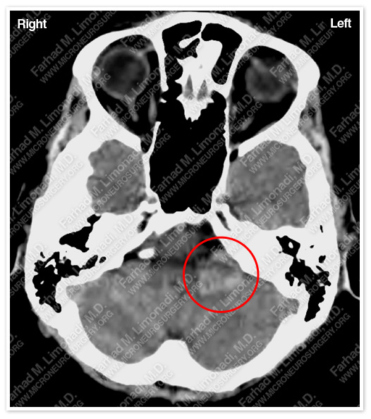
CT scan of patient’s brain shows a left hemorrhagic cerbellopontine angle tumor pressing on brain stem.
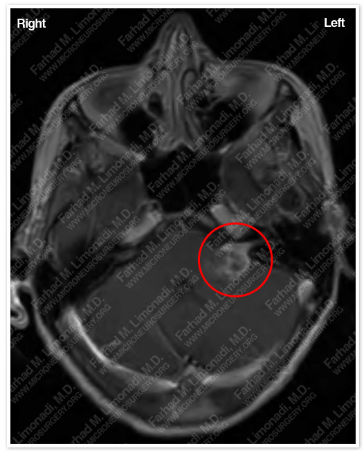
MRI scan of patient’s brain shows a left acoustic neuroma with central necrosis and hemorrhage.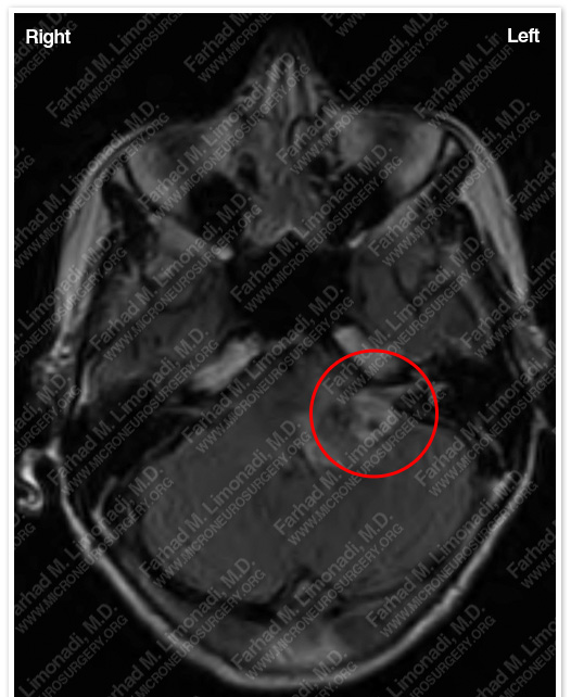
MRI scan flair sequence shows significant vasogenic edema involving brain stem and cerebellum.
Surgical Procedure
- She underwent a left translabyrinthine craniectomy and surgical resection of this brain tumor using intra-operative neurophysiological monitoring including facial nerve monitoring and brain stem auditory evoked response (BAER).
Pathology
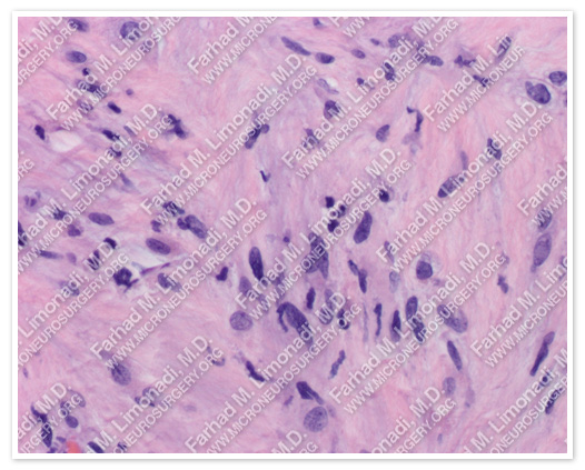
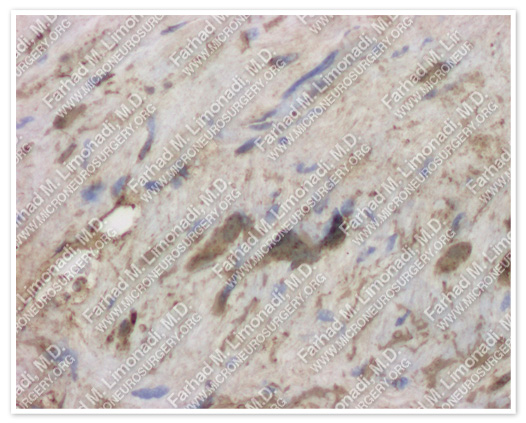
The pathology of the tumor confirmed diagnosis of acoustic neuroma (vestibular schwannoma).
Post-op Imaging
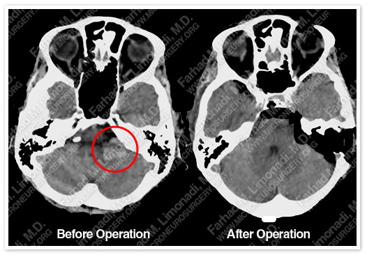
Post-op CT shows complete resection of the tumor with no injury to surrounding neurovascular structures.
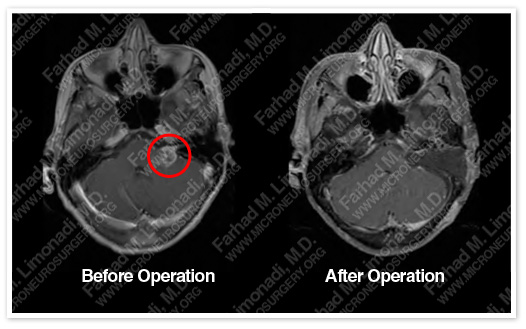

Post-op MRIs confirm complete resection of the tumor with no injury to surrounding neurovascular structures.
Post-op Course
- The patient did well postoperatively and was discharged with no additional neurological deficit. Her facial weakness and swallowing difficulty improved immediately; her facial numbness persisted for a few months and gradually improved.