


Case Presentation:
Metastatic Brain Tumors - Case 10
Metasatic Melanoma Brain Tumor
History and Physical
- 73-year-old gentleman with history of shoulder melanoma and recent diagnosis of metastatic melanoma of right upper lobe of lung, and status post successful resection of this tumor who presented with metastatic tumor (melanoma) to the brain.
- On examination, he had no focal neurological deficits.
Imaging
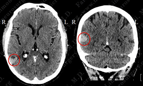
CT scan of the patient’s brain shows a tumor within posterior border of right temporal lobe of the brain.
Computer Navigation
- Stereotaxy and computer navigation was utilized to localize and remove the tumor precisely, with surrounding neurovascular structures mapped out and protected.
- Right temporal craniotomy approach was utilized.
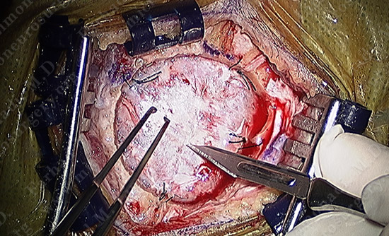
Small circular bone flap is elevated using computer localization. The dura (covering of the brain) is exposed.
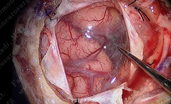
Dura is opened and the tumor which is stained dark is in the right upper corner of the exposure.
Pathology
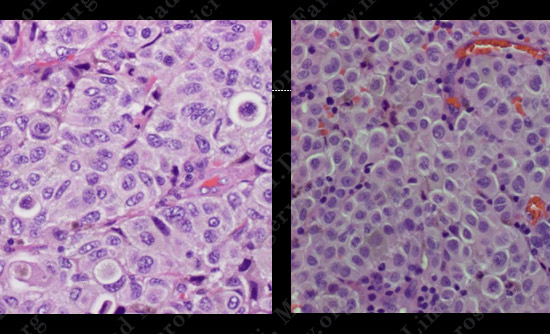
H&E slide of lung tumor H&E slide of brain tumor
The pathology of the tumor confirmed diagnosis of melanoma (similar to lung tumor specimen).
Post-op Course
- The patient did well postoperatively with no neurological deficit. He was discharged home and returned to baseline function.