


Case Presentation:
Metastatic Ovarian Cancer - Case 6
- 72-year-old right handed lady with diagnosis of ovarian carcinoma three years ago with stage IIIC, having undergone laparotomy, bilateral salpingo-oophorectomy, primary tumor resection, omentectomy, and diaphragmatic stripping, and small bowel resection, presented with a solitary metastatic brain tumor in the right parietal-occipital region. She initially chose stereotactic radiosurgery (radiation treatment) instead of surgery. Unfortunatley despite the radiation treatment her tumor grew in size and resulted in tonic-clonic seizure.
- On examination she had no focal neurological deficit and was completely alert and oriented.
Imaging
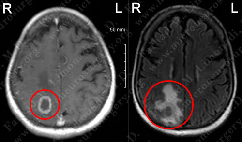
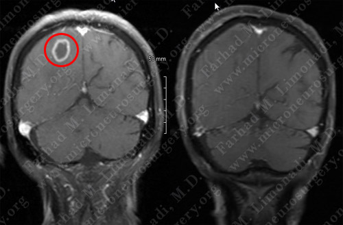
MRI scan of the patient's brain showed a right parietal-occipital brain tumor with ring enhancement (left) and significant vasogenic edema (right).
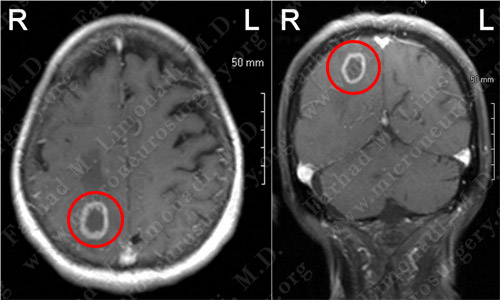
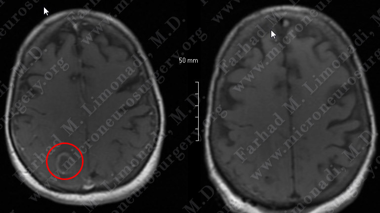
MRI scan of the patient's brain showed a right parietal-occipital brain tumor with ring enhancement on axial (left) and coronal view (right).
Computer Navigation
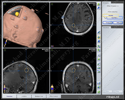
Computer navigation and stereotaxy utilized to map and localize the tumor (outlined in yellow) during surgery.
Surgical Procedure
- She underwent surgical resection of this tumor utilizing brain mapping, stereotactic and computer navigation, and intra-operative neurophysiological monitoring.
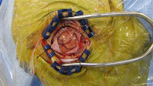
Utilizing computer navigation, a small craniotomy was performed precisely over the tumor and the tumor was removed using this small opening.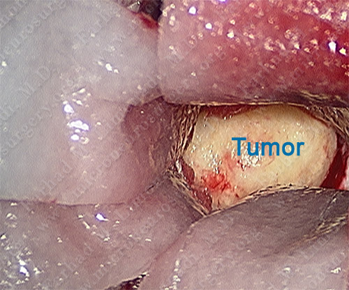
Tumor was isolated using this small opening.
Tumor was removed in a gross total en-bloc fashion
PathoIogy

The pathology of the tumor confirmed diagnosis of metastatic ovarian carcinoma.
Post-op Imaging
 Before Operation After Operation
Post-op MRI shows complete resection of the tumor with no injury to surrounding neurovascular structures.
Post-op Course
- The patient did well postoperatively with no neurological deficit. She remained seizure free and was discharged home.