


Case Presentation:
Metastatic Cervical Cancer - Case 12
Right Temporal Brain Tumor
History and Physical
- 30+ year old lady with headache, slow thought process, and confusion.
- On examination, she had no focal neurological deficit and was completely alert and oriented.
Imaging
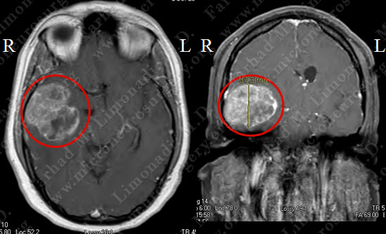
MRI scan of patient’s brain showed a right temporal brain tumor.
Computer Navigation
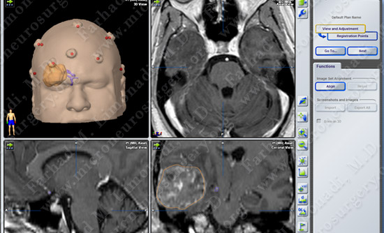
Stereotaxy and computer navigation was utilized to localize and remove the tumor precisely, with surrounding neurovascular structures mapped out.
Surgical Procedure
- She underwent surgical resection of this tumor utilizing brain mapping, stereotactic and computer navigation, and intraoperative neurophysiological monitoring.
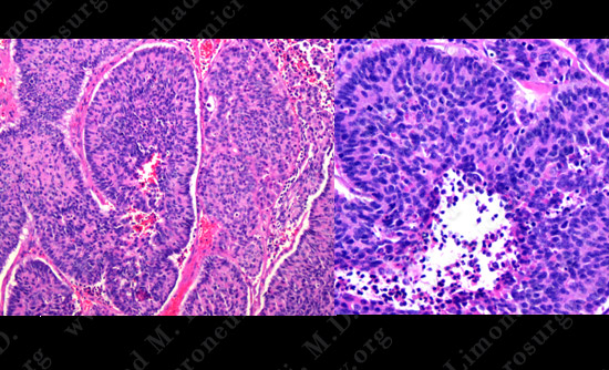
Pathology
Post-op MRI shows complete resection of the tumor with no injury to surrounding neuro-vascular structures.
Post-op Imaging
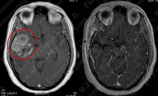
Before Operation After Operation
Post-op MRI shows complete resection of the tumor with no injury to surrounding neurovascular structures.
Post-op Course
- The patient did well postoperatively with no neurological deficit. She was discharged home and returned to normal function.