


Case Presentation:
Metastatic Brain Tumor - Case 2
History & Physical
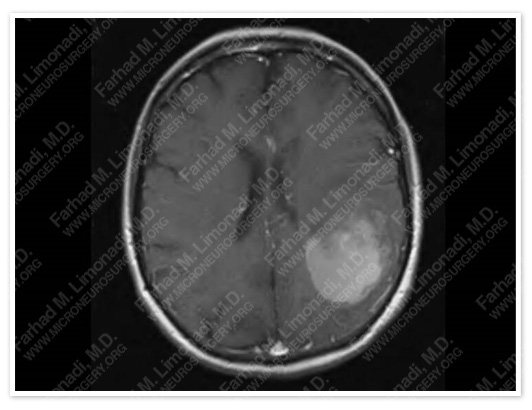
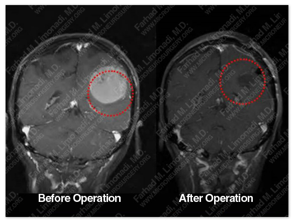
MRI scan of her brain showed a large tumor in the Right fronto-parietal cortex abutting the motor cortex.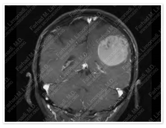
MRI scan of her brain showed a large tumor in the Right fronto-parietal cortex abutting the motor cortex.
- 65-year-old right handed lady with the history of metastatic ovarian cancer who presented with dysarthria and difficulty with bringing the right word out (anomia).
- Her condition rapidly evolved into inablility in moving her right arm and leg, and speak (aphasia, and right hemiparesis) and she was brought to the emergency room.
- Although patient could understand spoken language, she was unable to express herself (expressive aphasia).
Surgical Procedure
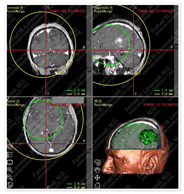
Stereotactic map of the brain was created with 1 mm accuracy around the lesion. The tumor was volumetrically mapped and removed after identifying and avoiding injury to motor cortex.
- She underwent image guided frameless stereotactic craniotomy with intraoperative motor cortex mapping.
Post op Course
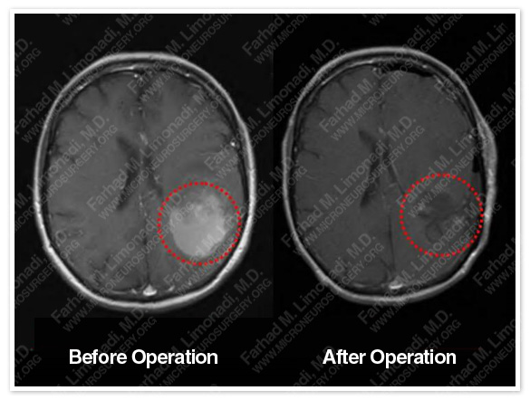

- Postoperatively her language function normalized. Further, her Right sided weakness immediately improved and she was able to walk and use her right arm at a normal level. She was discharged from the hospital.