


Case Presentation:
Metastatic Lung Cancer - Case 13
Left Occipital and Left CPA Brain Tumor
History and Physical
- 80-year-old gentleman with history of lung cancer three years ago treated with chemotherapy and radiotherapy, bladder cancer 8 years ago, and melanoma thirty years ago. He presented with difficulty with word recognition and reading.
- On examination, he had no focal neurological deficit and was completely alert and oriented.
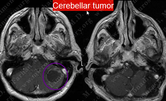
Imaging
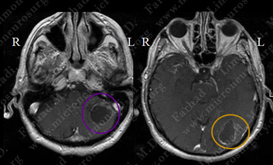
MRI scan of the patient’s brain showed a left suboccipital/cerebellar brain tumor (left image) and a left occipital tumor (right image). They are cystic with ring-enhancing signal.
Computer Navigation
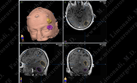
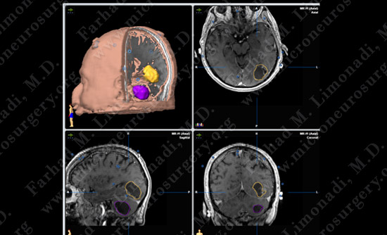
Stereotaxy and computer navigation was used intraoperatively to pinpoint the location of each tumor (outlined with purple and yellow).
Surgical Procedure
- He underwent surgical resection of both tumors utilizing brain mapping, stereotactic and computer navigation, and intraoperative neurophysiological monitoring.
- Computer navigation was utilized to localize each tumor with pinpoint accuracy.
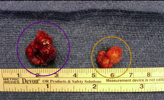
Both tumors were resected and were sent for pathological examination.
Pathology
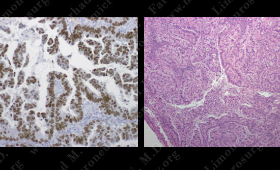
The pathology of the tumors confirmed diagnosis of metastatic pulmonary carcinoma.
Post-op Imaging
Post-op MRI shows complete resection of the tumor with no injury to surrounding neurovascular structures.