


Case Presentation:
Metastatic Brain Tumor - Case 11
Metastatic Melanoma
History and Physical
- 60+ year old lady presented with a history of lung cancer and metastatic tumor of right frontal brain. Her tumor was initially treated with stereotactic radiation treatment and followed radiographically. However, the tumor continued to grow despite radiation treatment and she was indicated for surgical resection of this tumor.
- On examination, she had no focal neurological deficit.
Imaging
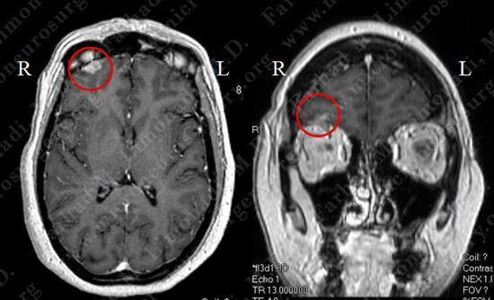
MRI scan of the patient’s brain showed the right frontal tumor above the orbit.
Computer Navigation
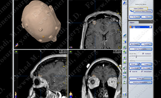
Stereotaxy and computer navigation was used intraoperatively to pinpoint the tumor (outlined in yellow).
Surgical Procedure
- She underwent surgical resection of the tumor using computer navigation and stereotaxy together with intraoperative neurophysiological monitoring.
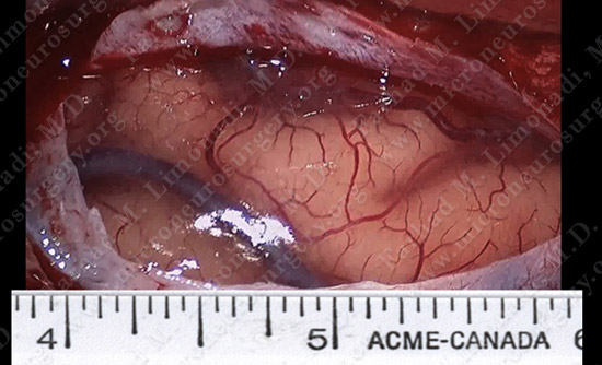
Surgical view of the right frontal lobe of the brain through the surgical microscope.
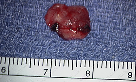
The tumor was resected and was sent for pathological evaluation.
Post-op Course
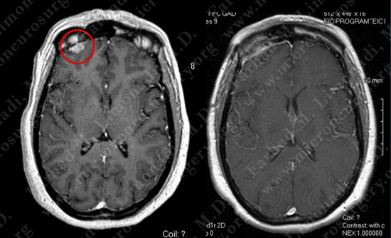
Before Operation After Operation
Post-op MRI shows complete resection of the tumor with no injury to surrounding neurovascular structures.