


Case Presentation:
Metastatic Ovarian Cancer - Case 7
History and Physical
- 65-year-old lady with history of metastatic ovarian cancer with initial diagnosis in 2006, status post chemotherapy, together with suboccipital craniectomy and resection of brain tumor in 2008 by a different neurosurgeon, with subsequent recurrence of the tumor which was initially managed by stereotactic radiosurgery at a different institution. Unfortunately, the tumor grew in size despite all this and she presented with recurrence and enlargement of this tumor.
- On examination, she had no focal neurological deficit and was completely alert and oriented.
Imaging
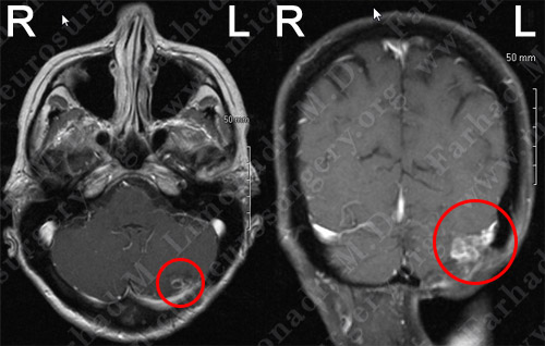
MRI scan of the patient’s brain showed a left suboccipital/cerebellar brain tumor.
Surgical Procedure
- She underwent surgical resection of this tumor utilizing brain mapping, stereotactic and computer navigation, and intraoperative neurophysiological monitoring. Her cosmetic deformity as the result of the craniectomy and radiation was also repaired with cranioplasty. The tumor was found to be growing in a large vascular bed of tentorium and dural sinuses.
PathoIogy
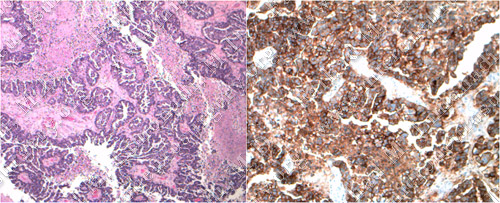
The pathology of the tumor confirmed diagnosis of metastatic ovarian carcinoma.
Post-op Imaging
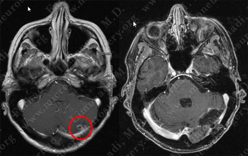
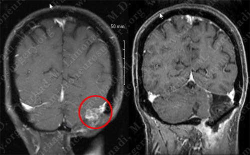
Before Operation After Operation
Post-op MRI shows complete resection of the tumor with no injury to surrounding neurovascular structures.
Post-op Course
- The patient did well postoperatively with no neurological deficit. She remained seizure free and was discharged home.