


Case Presentation:
Metastatic Brain Tumors - Case 8
Right Parietal-Occipital Brain Tumor
History and Physical
- 72-year-old right handed lady with known diagnosis of squamous lung cancer who presented with a brain tumor.
-
On examination, she had no focal neurological deficit except loss of vision in left visual field.
Imaging
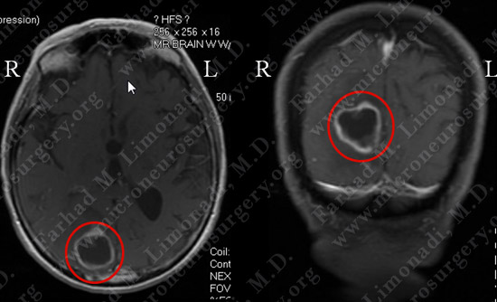
MRI scan of the patient’s brain showed a right parietal-occipital brain tumor with ring enhancement on axial (left) and coronal view (right).
Computer Navigation
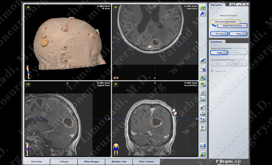
Computer navigation and stereotaxy utilized to map and localize the tumor (outlined in yellow) during surgery.
Surgical Procedure
- She underwent surgical resection of the tumor utilizing brain mapping, stereotactic and computer navigation, and intraoperative neurophysiological monitoring.
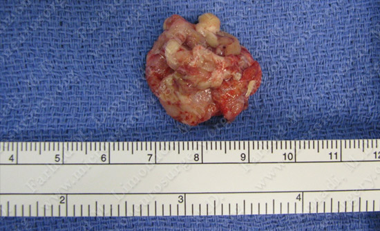
Tumor was removed in a gross total enbloc fashion.
PathoIogy
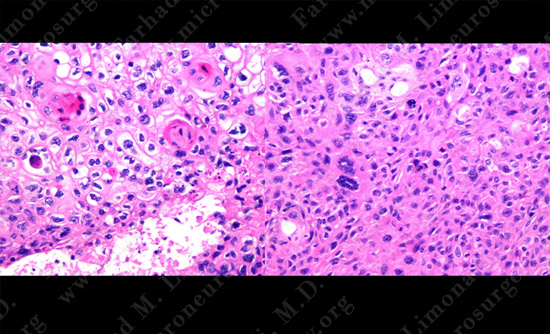
The pathology of the tumor confirmed diagnosis of metastatic squamous cell carcinoma.
Post-op Imaging
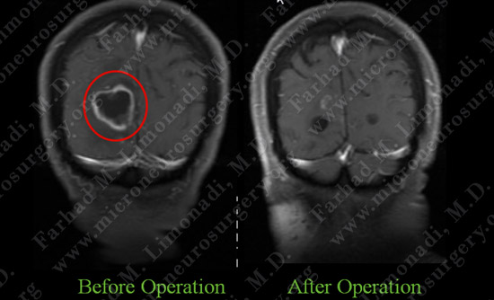
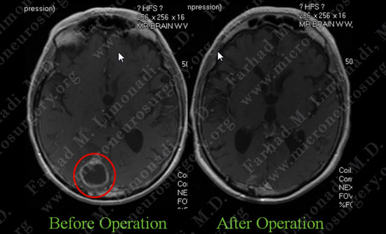
Post-op MRI shows complete resection of the tumor with no injury to surrounding neurovascular structures.
Post-op Course
- The patient did well postoperatively with no new neurological deficit. She was discharged home.