Case Presentation:
Metastatic Lung Cancer - Case 3
- 62-year-old lady who was admitted to the hospital with headache and dizziness. A comprehensive work-up showed primary lung cancer with 3 metastatic tumors in the brain.
- On examination, she had no focal neurological deficit. She had right finger to nose dysmetria.
Imaging
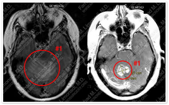
MRI scan of the patient’s brain shows three brain tumors. The largest tumor in the cerebellum is shown in the T1C sequence on left and flair sequence on right.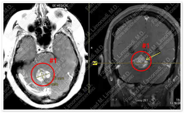
Additional views of this tumor show effacement of the fourth ventricle and mass effect on brain stem.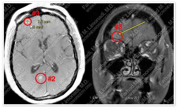
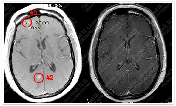
Second and third tumors are denoted as #2 and #3 in these two images.
Surgical Procedure
- She underwent surgical resection of the large cerebellar tumor, followed by stereotactic radiosurgery (SRS) to the other two tumors. She further underwent SRS to the cavity of the tumor resection site.
Post-op Imaging
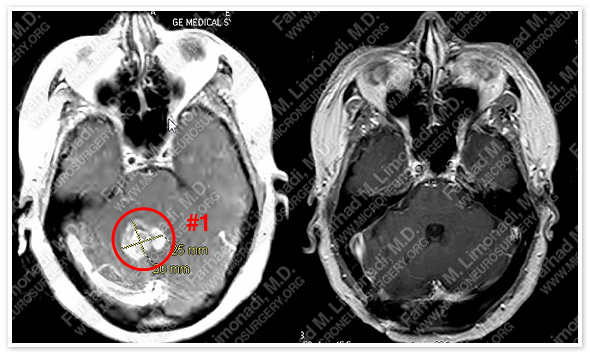 Before Operation After Operation
Post-op MRI shows complete resection of the large cerebellar tumor with no injury to surrounding neurovascular structures, and reopening of the fourth ventricle.
 Before Operation After Operation
Post-SRS MRI shows complete disappearance of the second and third tumors with no injury to surrounding neurovascular structures.


