


Case Presentation:
Meningioma - Case 1
History & Physical
- 67-year-old gentleman presented with confusion, decreased cognition, left-sided weakness, inability to walk, or use of his left hand.
Imaging
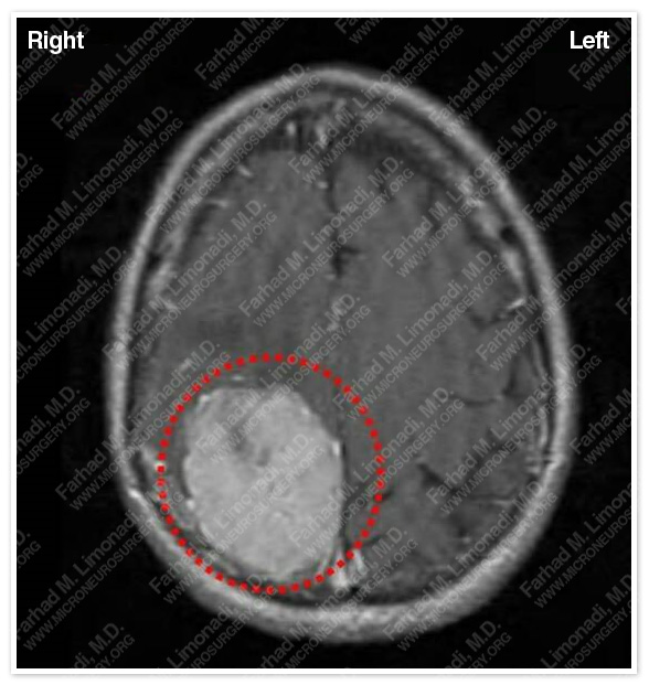
MRI scan of his brain showed a large right parieto-occipital tumor radiographically consistent with meningioma. Tumor is outlined in a red dotted circle.
Computer Navigation
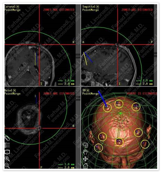
Comptuer navigation was used to determine the precise location of the tumor and important adjacent structure.
Surgical Procedure
- Patient underwent frameless stereotactic craniotomy and complete resection of the tumor.
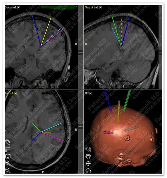
- Volumetric map of tumor together with neurophysiological cortical mapping assisted in safe and complete resection of the tumor while safeguarding vital neurovascular structures.
Post-op Imaging
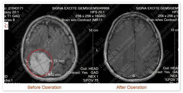
Post operative MRI showed complete resection of the tumor.
Post-op Course
- Patient was discharged home in a few days in good health and with no postsurgical complications. His symptoms caused by the tumor were completely resolved and he returned to normal function.