


Case Presentation:
Meningioma - Case 7
History & Physical
- 77-year-old right-handed man who presented with headache, blurry vision in the left eye, and focal seizure.
- On physical examination, he had decreased visual acuity in the right eye and decreased sensation to pin prick in the right upper and lower extremities.
Imaging
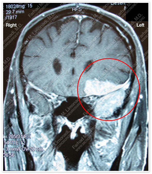
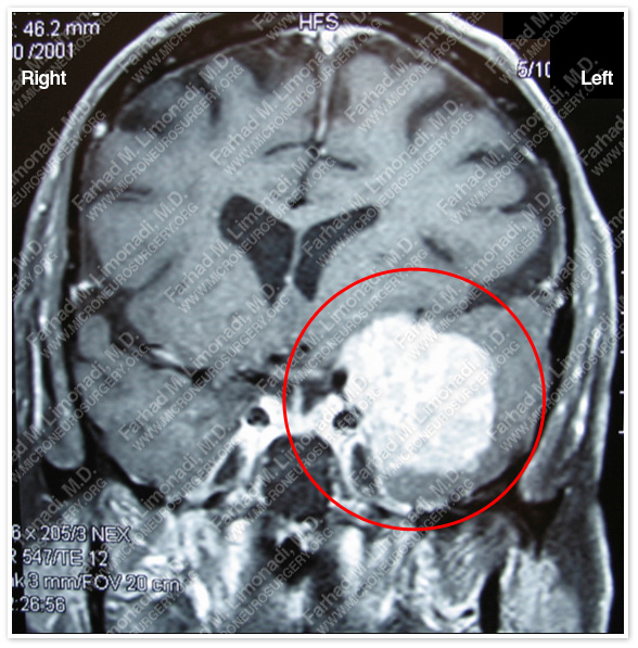
MRI scan of the patient’s brain shows a left extraaxial tumor at the sphenoid wing extending in the frontal and temporal lobe region.
Computer Navigation
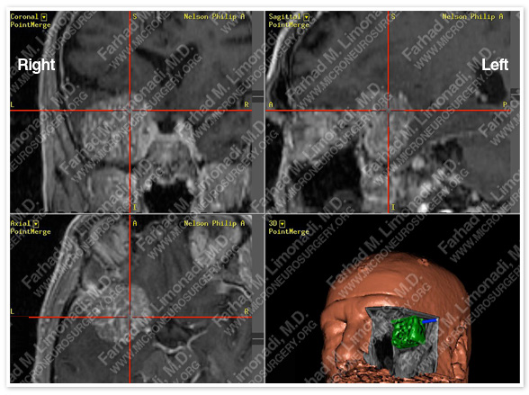
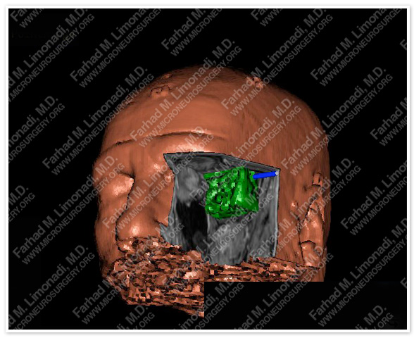
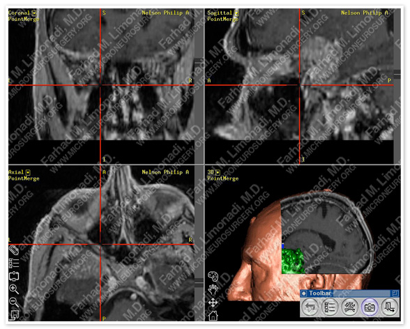
Computer simulation and modeling was utilized for precise localization of tumor (green in the right lower pic, and outlined in red circle).
Surgical Procedure
- He underwent orbitozygomatic craniotomy and surgical resection of this brain tumor using brain mapping, stereotactic and computer navigation, and intraoperative neurophysiological monitoring.
Post-op Imaging
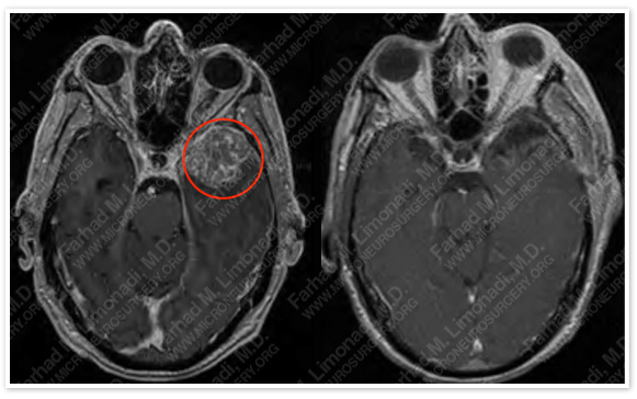
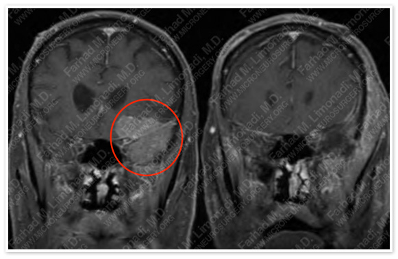
Before Operation After Operation
Post-op MRI shows complete resection of the tumor with no injury to surrounding neurovascular structures.
Post-Op Course
- The patient did well postoperatively and was discharged with no neurological deficit.