


Case Presentation:
Meningioma - Case 12
History & Physical
- 73-year-old gentleman who had undergone a subtotal resection of a complex dural based tumor found to be a meningioma over a year ago after presenting with aphasia (loss of speech), and seizure. His residual tumor was treated with stereotactic radiosurgery, however, despite this his tumor re-grew and resulted in seizure, aphasia (loss of speech), and hemiparesis (right-sided weakness).
-
His examination showed hemiparesis and aphasia. These symptoms improved with steroid administration temporarily.
Imaging
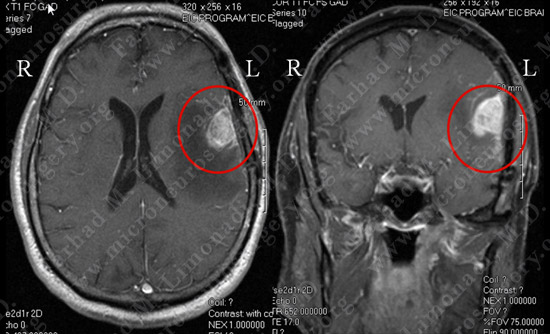
MRI scan of the patient’s brain shows a large dural based tumor in the left frontoparietal convexity overlying the speech cortex, with significant vasogenic edema.
Computer Navigation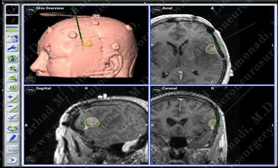
He underwent surgical resection of this tumor utilizing brain mapping, stereotactic and computer navigation, and intraoperative neurophysiological monitoring.
Surgical Procedure
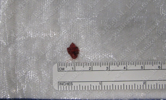
Tumor was removed in a gross total fashion.
Pathology
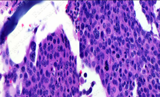
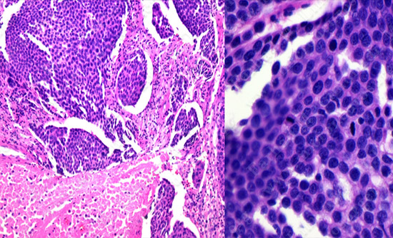
The pathology of the tumor confirmed diagnosis of meningioma.
Post-op Imaging
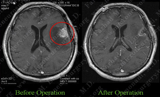
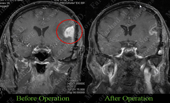
Post-op MRI shows complete resection of the tumor with no injury to surrounding neurovascular structures
Post-op Course
- Postoperatively, the patient returned to completely normal neurological status with no complications. He was discharged home.