


Case Presentation:
Occipital Meningioma - Case 17
History & Physical
- 55-year-old lady who presented with altered mentation, dysarthria, and hallucination.
- On examination, she had no focal neurological deficit and was completely alert and oriented.
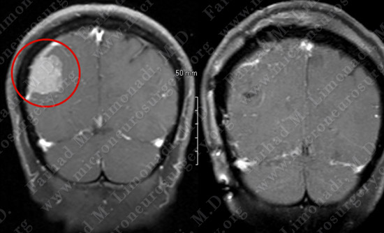
Imaging
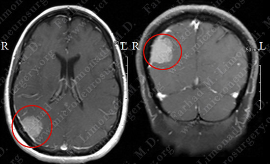
MRI scan of the patient’s brain showed a right occipital brain tumor.
Computer Navigation
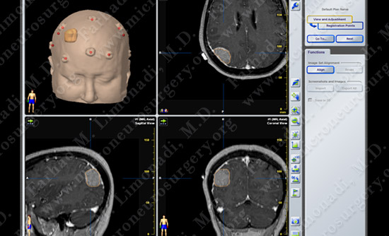
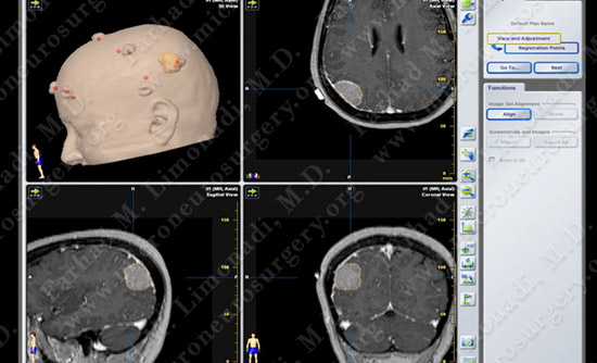
Computer navigation: Tumor is outlined in yellow.
Surgical Procedure
- She underwent right occipital craniotomy and resection of this tumor utilizing computer navigation and stereotaxy as well as intraoperative neurophysiological monitoring.
Pathology
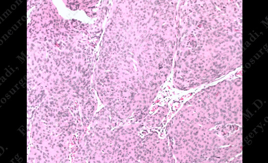
The pathology of the tumor confirmed the diagnosis of meningioma.
Post-op Imaging
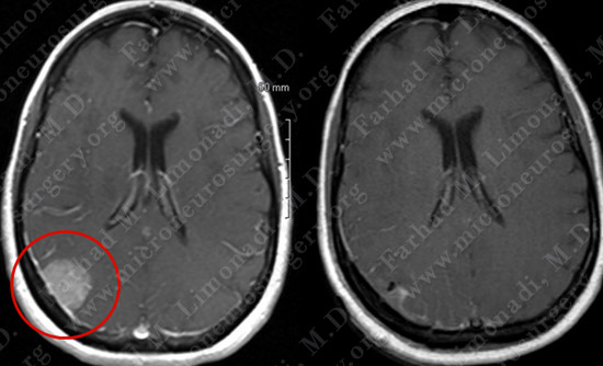
Before Operation After Operation
Before Operation After Operation
Post-op MRI shows complete resection of the tumor with no injury to surrounding neurovascular structures.
Post-op Course
- The patient did well postoperatively with no neurological deficit. She was discharged home with complete resolution of her preoperative symptoms. She returned to full time employment.