


Case Presentation:
Meningioma - Case 11
History & Physical
- 67-year-old right-handed lady who presented with headache of longstanding duration, however, worsening for the past six months.
- Patient examination showed generalized fatigue and otherwise she had a non focal examination.
Imaging
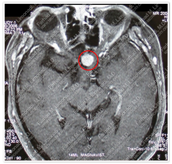
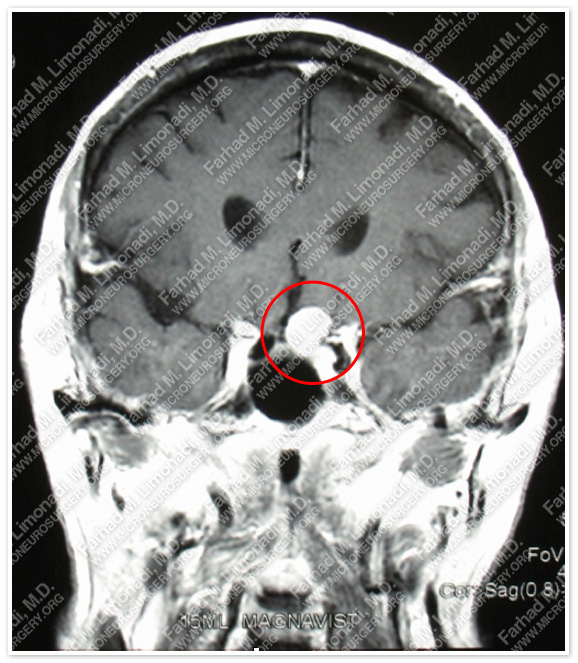
MRI scan of the patient’s brain shows a dural based tumor in the suprasellar, or tuberrculum sellae.
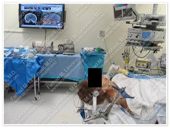
Surgical Procedure
- She underwent surgical resection of this tumor by supraorbital craniotomy and using brain mapping, stereotactic and computer navigation, and intraoperative neurophysiological monitoring.
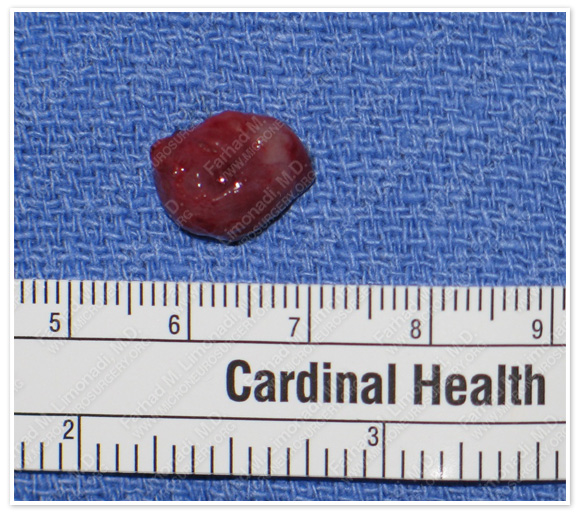
Tumor was removed in a gross total fashion.
Pathology
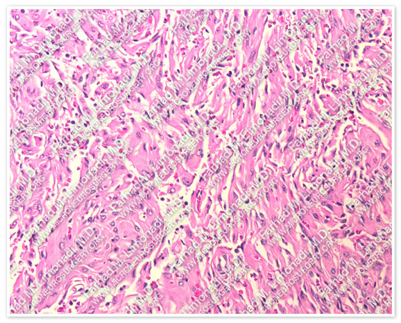
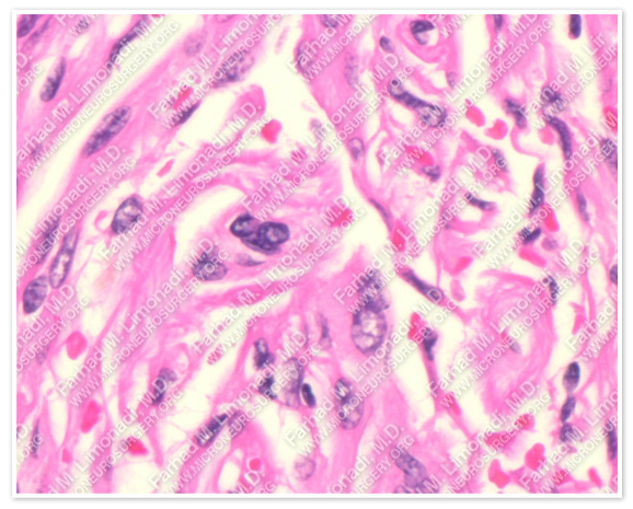
The pathology of the tumor confirmed diagnosis of meningioma.
Post-op Imaging
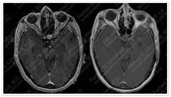
Post-op MRI shows complete resection of the tumor with no injury to surrounding neurovascular structures.
Post-op Course
- The patient did well postoperatively with no complications and was discharged home in good health.