


Case Presentation:
Meningioma - Case 16
History & Physical
- 69-year-old man with known diagnosis of meningioma and two previous attempts at removal of the tumor at a different institution. The tumor was not removed due its vascularity and the proximity to optic nerve, middle cerebral artery, and carotid artery.
- On examination, he had no focal neurological deficits.
Imaging
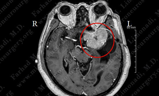
MRI scan of brain shows a left sphenoid wing meningioma arising from the dura (covering of brain) with intimate relationship with the optic nerve, middle cerebral artery, carotid artery. It is causing mass effect on the brainstem.
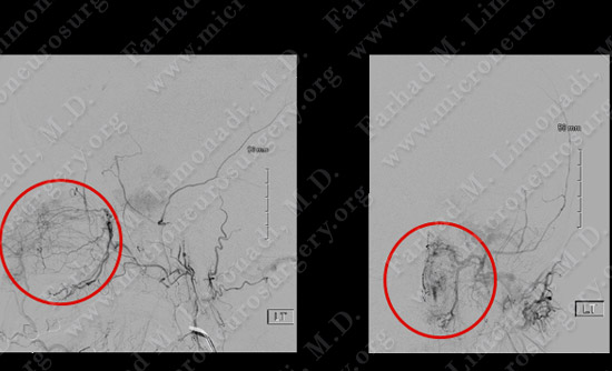
Vascular anatomy of the tumor was studies utilizing cerebral angiography. Tumor is outlined in red.
Computer Navigation
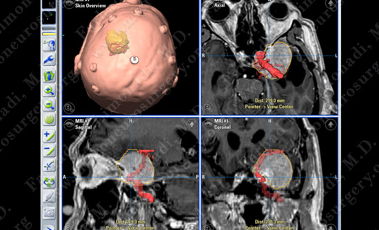
Computer navigation and stereotaxy was utilized to map out and localize the tumor (yellow) and critical vessels (red) intraoperatively.
Surgical Procedure
Post-op Imaging
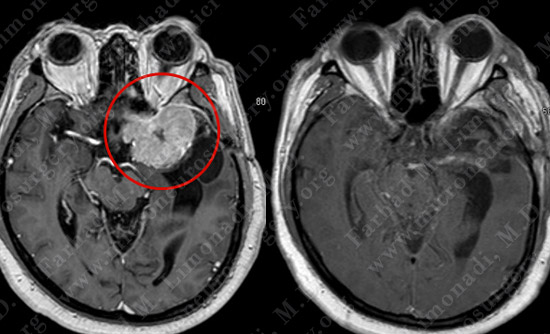
Before Operation After Operation
Post-op MRI shows complete resection of the tumor with no injury to surrounding neurovascular structures.