


Case Presentation:
Meningioma - Case 6
History & Physical
- 73-year-old gentleman who presented to the emergency room with headache, progressive loss of cognition and forgetfullness.
- On examination patient had no focal neurological deficit with exception of cognitive decline.
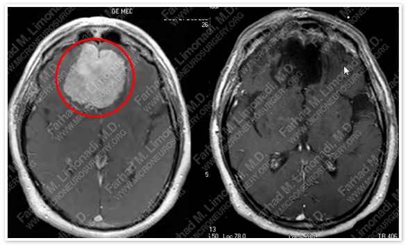
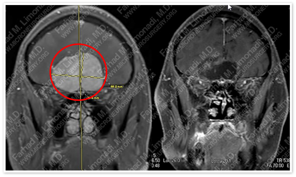
Imaging
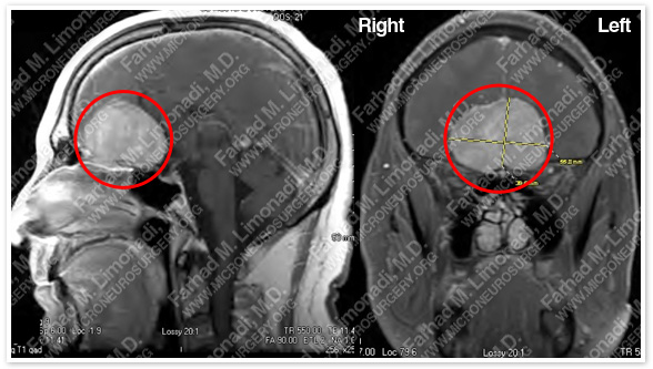
MRI scan of the patient’s brain shows a large olfactory groove tumor.
MRI shows significant vasogenic edema around this tumor that occupies a large volume of his brain.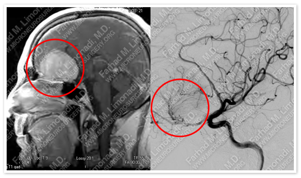
Cerebral angiography shows that this tumor is supplied by branches of the ophthalmic and anterior cerebral artery (right image).
Surgical Procedure
- He underwent bifrontal skull base craniotomy and resection of this tumor utilizing stereotactic and computer navigation and intraoperative neurophysiological monitoring.
Pathology
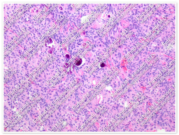
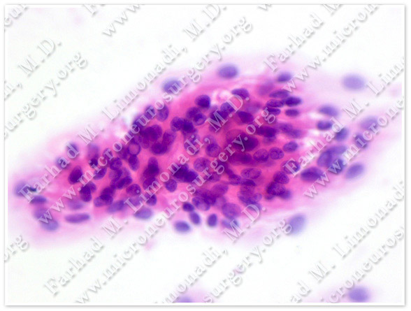
The pathology of the tumor confirmed diagnosis of olfactory groove meningioma.
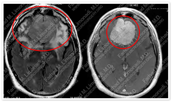
Post-op Imaging
Before Operation After Operation
Post-op MRI shows complete resection of the tumor with no injury to surrounding neurovascular structures.
Post-op Course
- The patient did well postoperatively despite having a prolonged hospitalization course due to postoperative edema, which was managed successfully. He was trached and pegged temporarily with ICP monitoring in the hospital, and was discharged from the hospital. He is tumor free and has returned to independent function.