


Case Presentation:
Occipital Meningioma - Case 19
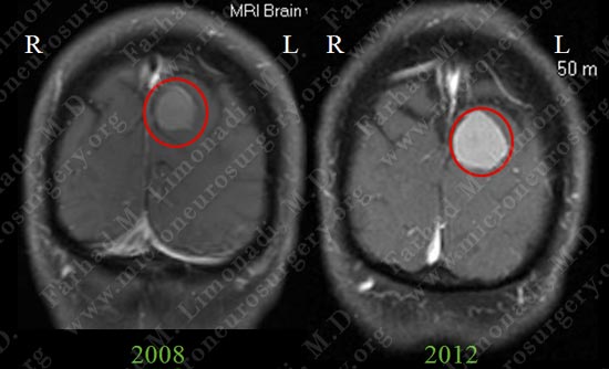
- 70-year-old lady with known diagnosis of an occipital meningioma which she had been completely asymptomatic for. She was followed radiographically and her tumor grew in size.
- On examination patient had no focal neurological deficit and had remained free of symptoms.
Imaging
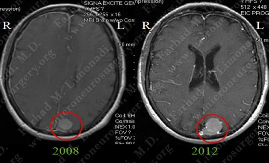
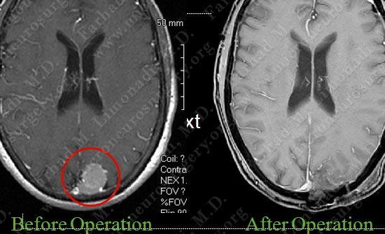
MRI scan of patient's brain shows tumor growth during 4 year followup. Tumor is encircled in red.
Surgical Procedure
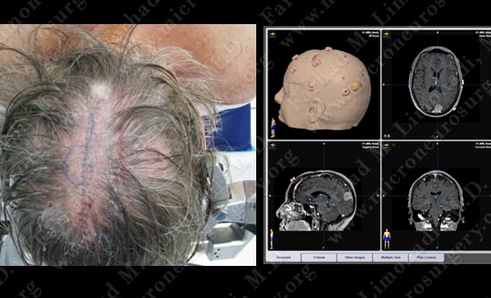
- She was a candidate for resection of this left occipital dural based tumor. After cardiac clearance, she was brought to the operating room and underwent stereotactic craniotomy and resection of the tumor utilizing stereotaxy/computer navigation and intra-operative neurophysiological monitoring.
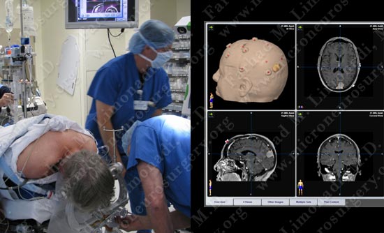
After patient has undergone general anesthesia, she is placed on the operating table in prone position and computer navigation is utilized to pinpoint the tumor.
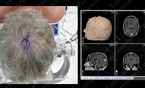
Computer navigation is utilized to pinpoint the tumor and its location is marked on the skin for planned incision and bony exposure.
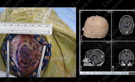
After the incision is made, computer navigation is again utilized to pinpoint the tumor and its location is marked on the skull for planned bony exposure.
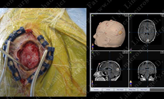
The bony exposure is precisely fashioned over the tumor. Here the dura is nicely exposed precisely over the tumor.
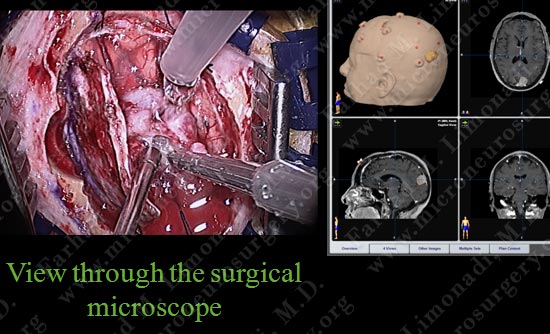
The tumor is removed utilizing ultrasonic aspirator (CUSA).
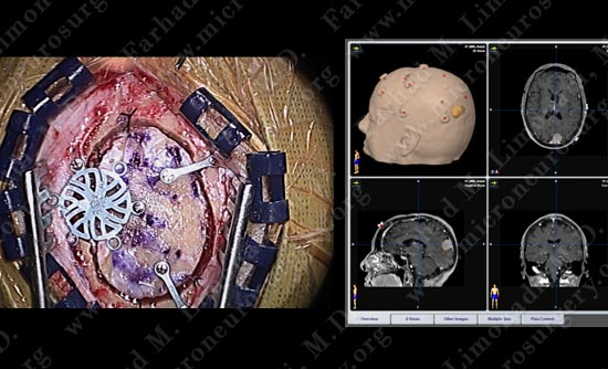
Following removal of the tumor and duraplasty, the bone is re-approximated using titanium low profile screws.
Scalp is re-approximated, patient is awakened and taken to the ICU for overnight observation.
Post-op MRI
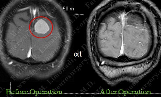
Post op MRI shows complete resection of the tumor with no injury to surrounding neuro-vascular structures.
- The patient did well post-operatively. She was discharged home in 3 days and returned to completely normal function.