


Case Presentation:
Meningioma, Petroclival - Case 13
History & Physical
-
63-year-old lady recovering from treatment of breast cancer, presented with right facial numbness, progressive difficulty with gait and balance, as well as light headedness.
-
On examination, she had no focal neurological deficit with exception of mild right facial droop and complete numbness in that region.
Imaging
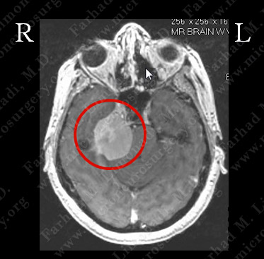
MRI scan of the patient’s brain showed a large right petro-clival meningioma with complete distortion of brain stem.
Computer Navigation
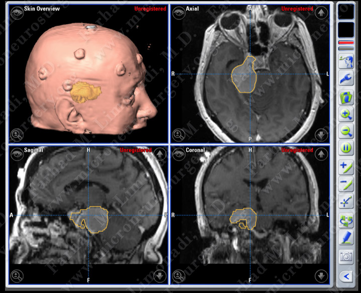
Computer navigation and stereotaxy were utilized to map and localize the tumor (outlined in yellow) during surgery. A retrosigmoid skull base approach was used to remove the infratentorial portion of this tumor with complete decompression of brainstem.
Surgical Procedure
-
After a period of conservative monitoring, the tumor was found to grow in size and subsequently she underwent a right temporal craniotomy and surgical resection of this tumor utilizing brain mapping, stereotactic and computer navigation, and intraoperative neurophysiological monitoring.
Pathology
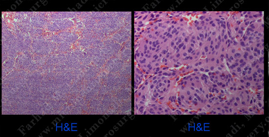
The pathology of the tumor revealed the diagnosis of meningioma.
Post-op Imaging
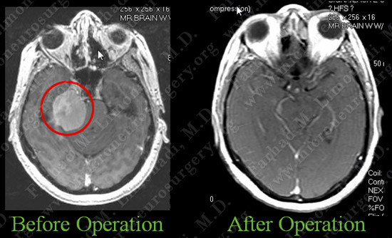
Post-op MRI shows radiographic resection of the infratentorial portion of the tumor with decompression of brainstem.
Post-op Course
- The patient did well postoperatively with no focal neurological deficit. Her facial sensation improved postoperatively.
.