


Case Presentation:
Meningioma - Case 2
Meningioma - Case 2
History & Physical
- 77-year-old man presented with new onset seizure
Imaging
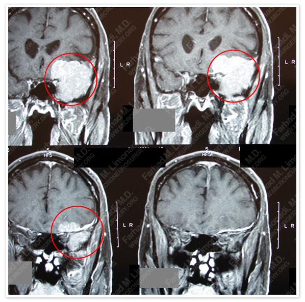
MRI scan of his brain showed a large left sphenoid wing tumor radiographically consistent with meningioma. Tumor is outlined in a red dotted circle.
Computer Navigation
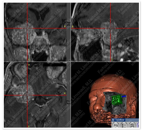
He underwent frameless stereotactic orbitozygomatic craniotomy and gross total resection of this tumor.
Post-op Imaging
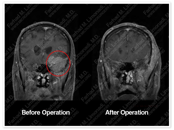
His post-op MRI showed complete resection of the tumor.
Post-op Course
- He was completelye intact neurologically and was discharged from the hospital a few days later.in good health.