


Case Presentation:
Occipital Meningioma - Case 18
History & Physical
- 30+ young lady who presented with severe headache of over 6 months duration.
- On physical examination she had a trace weakness on the right side of her body.
Imaging
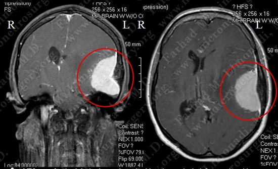
MRI scan of patient's brain shows a large dural based tumor in the left convexity with significant vasogenic edema and midline shift.
Surgical Procedure
- 30+ young lady who presented with severe headache of over 6 months duration.
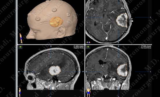
Stereotactic and computer navigation was used to determine the precise location of the tumor (outlined in yellow).
Pathology
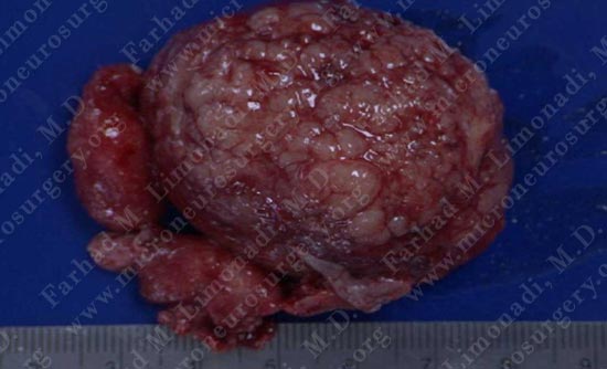
Tumor was removed in a gross total fashion.
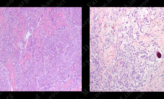
The pathology of the tumor confirmed diagnosis of meningioma.
Post-op Imaging
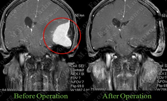
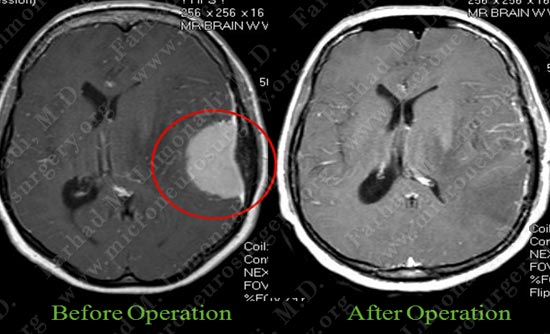
Post op MRI shows complete resection of the tumor with no injury to surrounding neuro-vascular structures
Post-op Course
- Post operatively patient returned to completely normal neurological status with no complications. He was discharged home.