


Case Presentation:
Meningioma - Case 8
History & Physical
- 77-year-old right-handed lady presented with a new onset of left-handed numbness. She was also suffering from dizziness, fatigue, and vertigo.
- On examination, patient had no focal finding.
Imaging
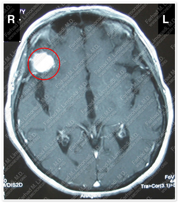
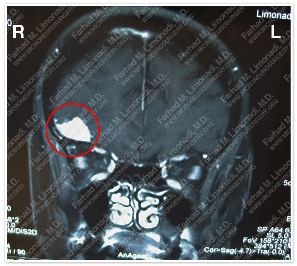
MRI scan of the patient’s brain shows a right extraaxial tumor at the sphenoid wing.
Surgical Procedure
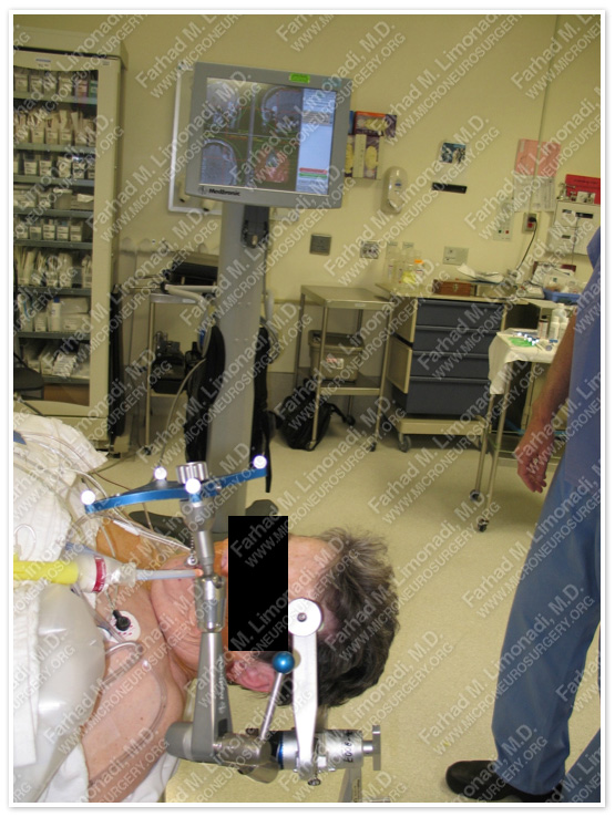
She underwent surgical resection of this tumor with the use of brain mapping, stereotactic and computer navigation, and intraoperative neurophysiological monitoring.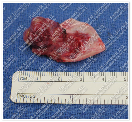
Tumor was removed in a gross total fashion. Tumor and its dural attachment are shown here.
Pathology
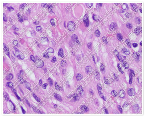
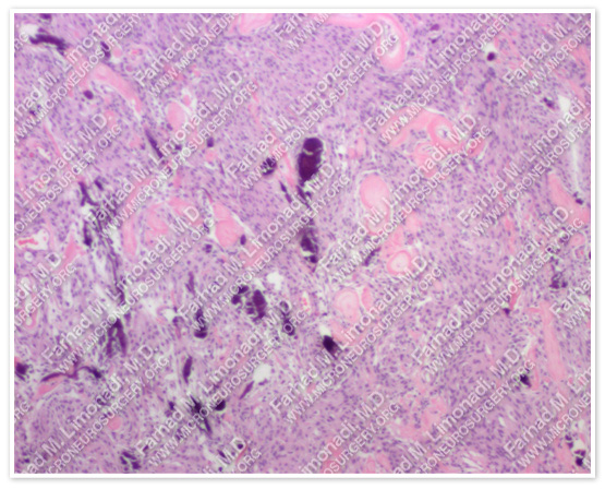
The pathology of the tumor confirmed diagnosis of meningioma
Post-op Imaging
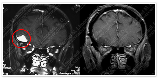
Before Operation After Operation
Post -p MRI shows complete resection of the tumor with no injury to surrounding neurovascular structures.
Post-op Course
- The patient did well post-operatively and was discharged with no neurological complication.