


Case Presentation:
Meningioma - Case 9
History & Physical
- 67-year-old left-handed lady who presented with a two-year history of progressive loss of visual acuity, double vision, and proptosis.
- Patient examination showed significant proptosis of her right eye, and significant loss of visual acuity.
Imaging
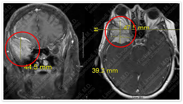
MRI scan of patient’s brain shows a sphenoid wing meningioma with intraconal extension.
Surgical Procedure
- She underwent surgical resection of this tumor by orbitozygomatic craniotomy and using brain mapping, stereotactic and computer navigation, and intraoperative neurophysiological monitoring.
Pathology
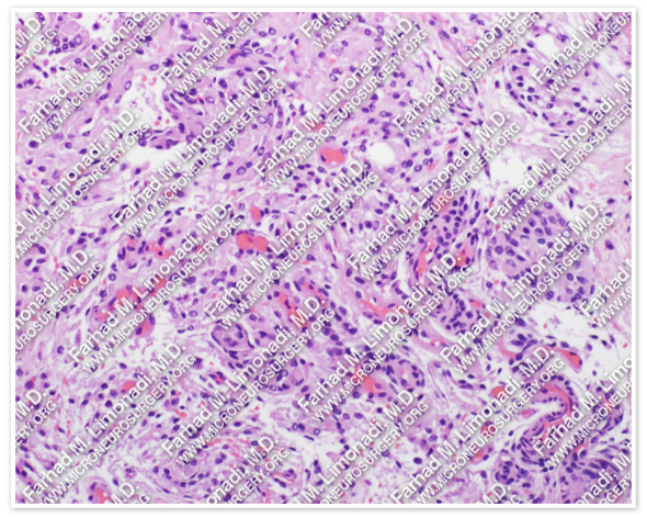
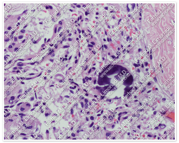
The pathology of the tumor confirmed diagnosis of meningioma.
Post-op Imaging
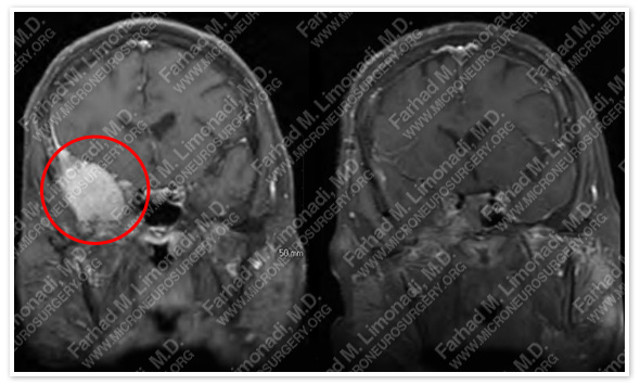
Before Operation After Operation
Post-op Course
- The patient did well postoperatively with no complications and was discharged home in good health.