


Case Presentation:
Meningioma - Case 5
History & Physical
- 70-year-old lady presented with right-sided dense hemiparesis (weakness) for about 2 weeks which culminated to a focal seizure.
- Patient examination showed significant right-sided weakness with strength of 2/5. She was unable to write, feed or groom herself.
Imaging
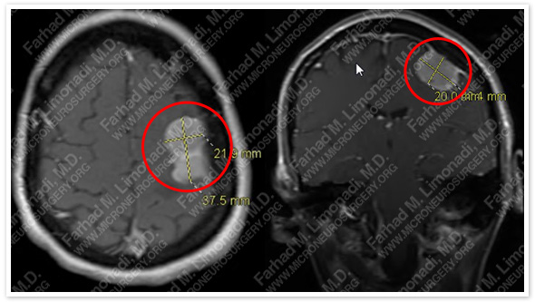
MRI scan of the patient’s brain shows a dural based tumor in the left frontoparietal convexity overlying the motor strip.
Computer Navigation
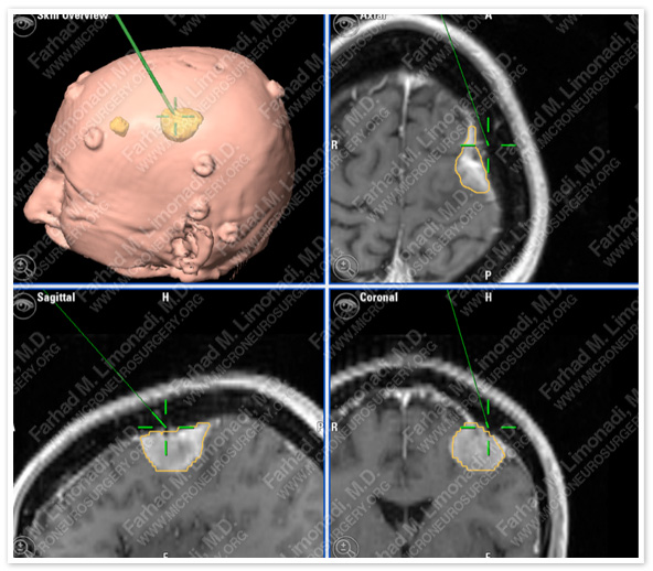
Stereotactic and computer navigation was used to determine the precise location of the tumor (outlined in yellow). She had a second benign tumor in front the symptomatic one which was treated conservatively as patient was not symptomatic with it.
Surgical Procedure
- She underwent surgical resection of this tumor utilizing brain mapping, stereotactic and computer navigation, and intraoperative neurophysiological monitoring.
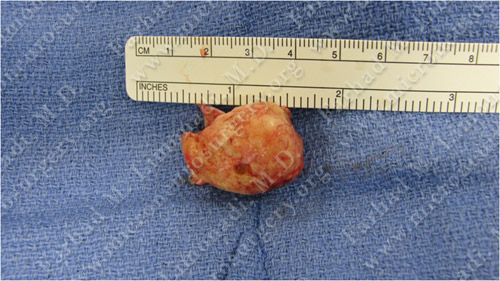
Tumor was removed in a gross total fashion.
Pathology
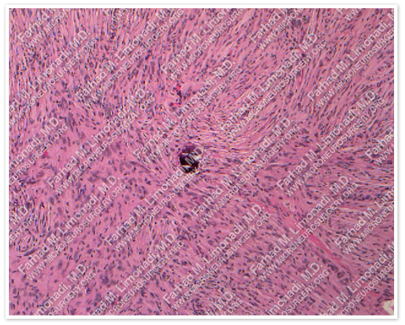
The pathology of the tumor confirmed diagnosis of meningioma.
Post-op Imaging
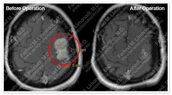
Post-op MRI shows complete resection of the tumor with no injury to surrounding neurovascular structures.