


Case Presentation:
Meningioma - Case 4
History & Physical
- 67-year-old man who presented to the emergency room with headache, nausea and vomiting, progressive difficulty with swallowing.
- On examination, the patient had no focal neurological deficit.
Imaging
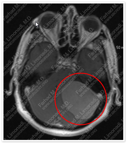
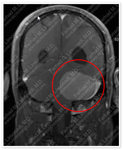
MRI scan of the patient’s brain shows a left extra-axial tumor at the cerebellopontine angle region, resulting in obstructive hydrocephalus with effacement of the fourth ventricle and mass effect on brainstem.
Surgical Procedure
- He underwent ventriculostomy followed by retro-sigmoid craniotomy and resection of this tumor utilizing stereotactic and computer navigation, intraoperative neurophysiological monitoring, including brainstem auditory evoked response (BAER), motor evoked potential (MEP), somatosensory evoked response (SSEP), and facial nerve monitoring.
Pathology
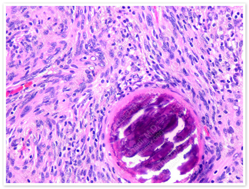
The pathology of the tumor confirmed diagnosis of transitional meningioma.
Post-op Imaging
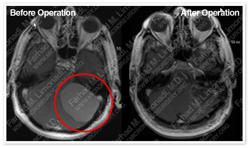
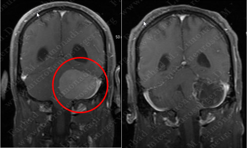
Post-op MRI shows complete resection of the tumor with no injury to surrounding neurovascular structures.